EP2492011A1 - Multiple laminar flow-based particle and cellular separation with laser steering - Google Patents
Multiple laminar flow-based particle and cellular separation with laser steering Download PDFInfo
- Publication number
- EP2492011A1 EP2492011A1 EP11193936A EP11193936A EP2492011A1 EP 2492011 A1 EP2492011 A1 EP 2492011A1 EP 11193936 A EP11193936 A EP 11193936A EP 11193936 A EP11193936 A EP 11193936A EP 2492011 A1 EP2492011 A1 EP 2492011A1
- Authority
- EP
- European Patent Office
- Prior art keywords
- flow
- components
- cells
- optical
- blood
- Prior art date
- Legal status (The legal status is an assumption and is not a legal conclusion. Google has not performed a legal analysis and makes no representation as to the accuracy of the status listed.)
- Withdrawn
Links
- 238000000926 separation method Methods 0.000 title claims abstract description 186
- 230000001413 cellular effect Effects 0.000 title abstract description 43
- 239000002245 particle Substances 0.000 title description 82
- 230000003287 optical effect Effects 0.000 claims abstract description 233
- 238000000034 method Methods 0.000 claims abstract description 133
- 238000000920 holographic laser trapping Methods 0.000 claims abstract description 27
- 210000004027 cell Anatomy 0.000 claims description 267
- 239000012530 fluid Substances 0.000 claims description 91
- 239000000463 material Substances 0.000 claims description 66
- 238000004062 sedimentation Methods 0.000 claims description 56
- 239000000203 mixture Substances 0.000 claims description 44
- 230000008569 process Effects 0.000 claims description 29
- 239000002699 waste material Substances 0.000 claims description 18
- 239000000126 substance Substances 0.000 claims description 17
- 239000000872 buffer Substances 0.000 claims description 16
- 239000011324 bead Substances 0.000 claims description 14
- 238000003384 imaging method Methods 0.000 claims description 14
- 238000005194 fractionation Methods 0.000 claims description 12
- 238000005259 measurement Methods 0.000 claims description 12
- 238000004590 computer program Methods 0.000 claims description 11
- 238000004611 spectroscopical analysis Methods 0.000 claims description 9
- 239000000470 constituent Substances 0.000 claims description 8
- 201000010099 disease Diseases 0.000 claims description 8
- 208000037265 diseases, disorders, signs and symptoms Diseases 0.000 claims description 8
- 210000000582 semen Anatomy 0.000 claims description 8
- 238000006243 chemical reaction Methods 0.000 claims description 7
- 238000001514 detection method Methods 0.000 claims description 7
- 230000002147 killing effect Effects 0.000 claims description 7
- 210000001766 X chromosome Anatomy 0.000 claims description 5
- 210000002593 Y chromosome Anatomy 0.000 claims description 5
- 239000007853 buffer solution Substances 0.000 claims description 5
- 238000001228 spectrum Methods 0.000 claims description 4
- 238000007689 inspection Methods 0.000 claims description 3
- 238000012632 fluorescent imaging Methods 0.000 claims description 2
- 230000000007 visual effect Effects 0.000 claims description 2
- 231100000518 lethal Toxicity 0.000 claims 2
- 230000001665 lethal effect Effects 0.000 claims 2
- 230000003213 activating effect Effects 0.000 claims 1
- 238000004520 electroporation Methods 0.000 claims 1
- 238000010183 spectrum analysis Methods 0.000 claims 1
- 239000000306 component Substances 0.000 abstract description 253
- 210000004369 blood Anatomy 0.000 abstract description 121
- 239000008280 blood Substances 0.000 abstract description 121
- 239000012503 blood component Substances 0.000 abstract description 34
- 238000000651 laser trapping Methods 0.000 abstract description 24
- 239000000243 solution Substances 0.000 description 58
- 210000001772 blood platelet Anatomy 0.000 description 53
- 239000000523 sample Substances 0.000 description 45
- 210000000265 leukocyte Anatomy 0.000 description 42
- 210000003743 erythrocyte Anatomy 0.000 description 37
- 238000000059 patterning Methods 0.000 description 26
- 239000002609 medium Substances 0.000 description 24
- 210000002381 plasma Anatomy 0.000 description 24
- 230000005684 electric field Effects 0.000 description 20
- 230000008901 benefit Effects 0.000 description 17
- 238000010586 diagram Methods 0.000 description 15
- 239000000047 product Substances 0.000 description 15
- 230000033001 locomotion Effects 0.000 description 14
- 230000004899 motility Effects 0.000 description 14
- 239000013049 sediment Substances 0.000 description 14
- 241000894006 Bacteria Species 0.000 description 13
- 239000000356 contaminant Substances 0.000 description 13
- 238000002156 mixing Methods 0.000 description 12
- 238000005406 washing Methods 0.000 description 12
- 241000700605 Viruses Species 0.000 description 11
- 239000003146 anticoagulant agent Substances 0.000 description 11
- 229940127219 anticoagulant drug Drugs 0.000 description 11
- 210000000601 blood cell Anatomy 0.000 description 11
- 230000005855 radiation Effects 0.000 description 11
- 238000010521 absorption reaction Methods 0.000 description 10
- 239000012620 biological material Substances 0.000 description 10
- 230000006870 function Effects 0.000 description 10
- 239000004973 liquid crystal related substance Substances 0.000 description 10
- 230000035899 viability Effects 0.000 description 10
- 239000003795 chemical substances by application Substances 0.000 description 9
- 230000007246 mechanism Effects 0.000 description 9
- 230000003068 static effect Effects 0.000 description 9
- 238000013459 approach Methods 0.000 description 8
- 238000005119 centrifugation Methods 0.000 description 8
- 238000009792 diffusion process Methods 0.000 description 8
- 238000005516 engineering process Methods 0.000 description 8
- 238000004519 manufacturing process Methods 0.000 description 8
- 238000012545 processing Methods 0.000 description 8
- 238000000746 purification Methods 0.000 description 8
- 238000003491 array Methods 0.000 description 7
- 238000013461 design Methods 0.000 description 7
- 238000001914 filtration Methods 0.000 description 7
- 229920000435 poly(dimethylsiloxane) Polymers 0.000 description 7
- 238000012546 transfer Methods 0.000 description 7
- VYPSYNLAJGMNEJ-UHFFFAOYSA-N Silicium dioxide Chemical compound O=[Si]=O VYPSYNLAJGMNEJ-UHFFFAOYSA-N 0.000 description 6
- 230000008859 change Effects 0.000 description 6
- 230000007423 decrease Effects 0.000 description 6
- 230000000694 effects Effects 0.000 description 6
- 239000000499 gel Substances 0.000 description 6
- 238000012544 monitoring process Methods 0.000 description 6
- 238000012576 optical tweezer Methods 0.000 description 6
- 210000003463 organelle Anatomy 0.000 description 6
- 230000008855 peristalsis Effects 0.000 description 6
- 230000002572 peristaltic effect Effects 0.000 description 6
- 238000009987 spinning Methods 0.000 description 6
- XLYOFNOQVPJJNP-UHFFFAOYSA-N water Substances O XLYOFNOQVPJJNP-UHFFFAOYSA-N 0.000 description 6
- 239000000084 colloidal system Substances 0.000 description 5
- 238000011109 contamination Methods 0.000 description 5
- 238000009826 distribution Methods 0.000 description 5
- 235000012489 doughnuts Nutrition 0.000 description 5
- 238000005286 illumination Methods 0.000 description 5
- 210000004153 islets of langerhan Anatomy 0.000 description 5
- 210000000947 motile cell Anatomy 0.000 description 5
- 230000000704 physical effect Effects 0.000 description 5
- 238000005086 pumping Methods 0.000 description 5
- 230000004044 response Effects 0.000 description 5
- 239000007787 solid Substances 0.000 description 5
- 210000001519 tissue Anatomy 0.000 description 5
- PEDCQBHIVMGVHV-UHFFFAOYSA-N Glycerine Chemical compound OCC(O)CO PEDCQBHIVMGVHV-UHFFFAOYSA-N 0.000 description 4
- XEEYBQQBJWHFJM-UHFFFAOYSA-N Iron Chemical compound [Fe] XEEYBQQBJWHFJM-UHFFFAOYSA-N 0.000 description 4
- 238000003556 assay Methods 0.000 description 4
- 238000010261 blood fractionation Methods 0.000 description 4
- 238000004720 dielectrophoresis Methods 0.000 description 4
- 239000000975 dye Substances 0.000 description 4
- 230000007613 environmental effect Effects 0.000 description 4
- 238000002474 experimental method Methods 0.000 description 4
- 238000000605 extraction Methods 0.000 description 4
- 235000013305 food Nutrition 0.000 description 4
- 230000005484 gravity Effects 0.000 description 4
- 230000003993 interaction Effects 0.000 description 4
- 150000002632 lipids Chemical class 0.000 description 4
- 239000012528 membrane Substances 0.000 description 4
- 102000004169 proteins and genes Human genes 0.000 description 4
- 108090000623 proteins and genes Proteins 0.000 description 4
- 150000003839 salts Chemical class 0.000 description 4
- 230000003595 spectral effect Effects 0.000 description 4
- 230000001954 sterilising effect Effects 0.000 description 4
- 238000004659 sterilization and disinfection Methods 0.000 description 4
- 239000000725 suspension Substances 0.000 description 4
- 206010053567 Coagulopathies Diseases 0.000 description 3
- XUIMIQQOPSSXEZ-UHFFFAOYSA-N Silicon Chemical compound [Si] XUIMIQQOPSSXEZ-UHFFFAOYSA-N 0.000 description 3
- 230000009471 action Effects 0.000 description 3
- 230000002776 aggregation Effects 0.000 description 3
- 238000004220 aggregation Methods 0.000 description 3
- 239000012472 biological sample Substances 0.000 description 3
- 230000005540 biological transmission Effects 0.000 description 3
- 210000001109 blastomere Anatomy 0.000 description 3
- 230000003139 buffering effect Effects 0.000 description 3
- 210000003850 cellular structure Anatomy 0.000 description 3
- 230000035602 clotting Effects 0.000 description 3
- 238000007796 conventional method Methods 0.000 description 3
- 230000003247 decreasing effect Effects 0.000 description 3
- 230000001066 destructive effect Effects 0.000 description 3
- 238000011161 development Methods 0.000 description 3
- 230000018109 developmental process Effects 0.000 description 3
- 238000000502 dialysis Methods 0.000 description 3
- 239000003085 diluting agent Substances 0.000 description 3
- 239000011521 glass Substances 0.000 description 3
- 238000011835 investigation Methods 0.000 description 3
- 230000004048 modification Effects 0.000 description 3
- 238000012986 modification Methods 0.000 description 3
- 230000037452 priming Effects 0.000 description 3
- 230000010349 pulsation Effects 0.000 description 3
- 230000003134 recirculating effect Effects 0.000 description 3
- 230000009467 reduction Effects 0.000 description 3
- 239000012488 sample solution Substances 0.000 description 3
- 238000001878 scanning electron micrograph Methods 0.000 description 3
- 229910052710 silicon Inorganic materials 0.000 description 3
- 239000010703 silicon Substances 0.000 description 3
- 239000000377 silicon dioxide Substances 0.000 description 3
- 241000894007 species Species 0.000 description 3
- 239000000758 substrate Substances 0.000 description 3
- 239000004094 surface-active agent Substances 0.000 description 3
- 238000011282 treatment Methods 0.000 description 3
- 241000283690 Bos taurus Species 0.000 description 2
- 241001465754 Metazoa Species 0.000 description 2
- 239000004988 Nematic liquid crystal Substances 0.000 description 2
- 206010028980 Neoplasm Diseases 0.000 description 2
- 240000004808 Saccharomyces cerevisiae Species 0.000 description 2
- FAPWRFPIFSIZLT-UHFFFAOYSA-M Sodium chloride Chemical compound [Na+].[Cl-] FAPWRFPIFSIZLT-UHFFFAOYSA-M 0.000 description 2
- 239000000654 additive Substances 0.000 description 2
- 239000000427 antigen Substances 0.000 description 2
- 102000036639 antigens Human genes 0.000 description 2
- 108091007433 antigens Proteins 0.000 description 2
- 230000004888 barrier function Effects 0.000 description 2
- 230000008512 biological response Effects 0.000 description 2
- 238000001574 biopsy Methods 0.000 description 2
- 238000004364 calculation method Methods 0.000 description 2
- 201000011510 cancer Diseases 0.000 description 2
- 238000012512 characterization method Methods 0.000 description 2
- 239000012141 concentrate Substances 0.000 description 2
- 238000010276 construction Methods 0.000 description 2
- 238000010924 continuous production Methods 0.000 description 2
- 229910003460 diamond Inorganic materials 0.000 description 2
- 239000010432 diamond Substances 0.000 description 2
- 238000007865 diluting Methods 0.000 description 2
- 238000010790 dilution Methods 0.000 description 2
- 239000012895 dilution Substances 0.000 description 2
- 235000013601 eggs Nutrition 0.000 description 2
- 238000001962 electrophoresis Methods 0.000 description 2
- 239000000284 extract Substances 0.000 description 2
- 230000008713 feedback mechanism Effects 0.000 description 2
- 239000012467 final product Substances 0.000 description 2
- 238000001943 fluorescence-activated cell sorting Methods 0.000 description 2
- 235000011187 glycerol Nutrition 0.000 description 2
- 238000009499 grossing Methods 0.000 description 2
- 230000000977 initiatory effect Effects 0.000 description 2
- 229910052742 iron Inorganic materials 0.000 description 2
- 210000003734 kidney Anatomy 0.000 description 2
- 150000002605 large molecules Chemical class 0.000 description 2
- 208000032839 leukemia Diseases 0.000 description 2
- 230000000670 limiting effect Effects 0.000 description 2
- 239000007788 liquid Substances 0.000 description 2
- 229920002521 macromolecule Polymers 0.000 description 2
- 238000007885 magnetic separation Methods 0.000 description 2
- 239000002184 metal Substances 0.000 description 2
- 229910052751 metal Inorganic materials 0.000 description 2
- 244000005700 microbiome Species 0.000 description 2
- 210000004940 nucleus Anatomy 0.000 description 2
- 239000013307 optical fiber Substances 0.000 description 2
- 230000003204 osmotic effect Effects 0.000 description 2
- 244000052769 pathogen Species 0.000 description 2
- 229920000642 polymer Polymers 0.000 description 2
- 239000002243 precursor Substances 0.000 description 2
- 238000003672 processing method Methods 0.000 description 2
- 230000002062 proliferating effect Effects 0.000 description 2
- 210000001747 pupil Anatomy 0.000 description 2
- 238000011160 research Methods 0.000 description 2
- 238000007493 shaping process Methods 0.000 description 2
- 230000035939 shock Effects 0.000 description 2
- 208000007056 sickle cell anemia Diseases 0.000 description 2
- 239000011780 sodium chloride Substances 0.000 description 2
- NLJMYIDDQXHKNR-UHFFFAOYSA-K sodium citrate Chemical compound O.O.[Na+].[Na+].[Na+].[O-]C(=O)CC(O)(CC([O-])=O)C([O-])=O NLJMYIDDQXHKNR-UHFFFAOYSA-K 0.000 description 2
- 239000001509 sodium citrate Substances 0.000 description 2
- 238000003860 storage Methods 0.000 description 2
- 230000009182 swimming Effects 0.000 description 2
- 230000009466 transformation Effects 0.000 description 2
- 238000000844 transformation Methods 0.000 description 2
- 239000012780 transparent material Substances 0.000 description 2
- 238000011144 upstream manufacturing Methods 0.000 description 2
- 230000003612 virological effect Effects 0.000 description 2
- 238000011179 visual inspection Methods 0.000 description 2
- 208000030507 AIDS Diseases 0.000 description 1
- 230000005653 Brownian motion process Effects 0.000 description 1
- 102000009123 Fibrin Human genes 0.000 description 1
- 108010073385 Fibrin Proteins 0.000 description 1
- BWGVNKXGVNDBDI-UHFFFAOYSA-N Fibrin monomer Chemical compound CNC(=O)CNC(=O)CN BWGVNKXGVNDBDI-UHFFFAOYSA-N 0.000 description 1
- HTTJABKRGRZYRN-UHFFFAOYSA-N Heparin Chemical compound OC1C(NC(=O)C)C(O)OC(COS(O)(=O)=O)C1OC1C(OS(O)(=O)=O)C(O)C(OC2C(C(OS(O)(=O)=O)C(OC3C(C(O)C(O)C(O3)C(O)=O)OS(O)(=O)=O)C(CO)O2)NS(O)(=O)=O)C(C(O)=O)O1 HTTJABKRGRZYRN-UHFFFAOYSA-N 0.000 description 1
- 102000029749 Microtubule Human genes 0.000 description 1
- 108091022875 Microtubule Proteins 0.000 description 1
- 108091034117 Oligonucleotide Proteins 0.000 description 1
- 239000004793 Polystyrene Substances 0.000 description 1
- 102000007056 Recombinant Fusion Proteins Human genes 0.000 description 1
- 108010008281 Recombinant Fusion Proteins Proteins 0.000 description 1
- 241000270295 Serpentes Species 0.000 description 1
- 210000001744 T-lymphocyte Anatomy 0.000 description 1
- 208000019513 White blood cell disease Diseases 0.000 description 1
- JLCPHMBAVCMARE-UHFFFAOYSA-N [3-[[3-[[3-[[3-[[3-[[3-[[3-[[3-[[3-[[3-[[3-[[5-(2-amino-6-oxo-1H-purin-9-yl)-3-[[3-[[3-[[3-[[3-[[3-[[5-(2-amino-6-oxo-1H-purin-9-yl)-3-[[5-(2-amino-6-oxo-1H-purin-9-yl)-3-hydroxyoxolan-2-yl]methoxy-hydroxyphosphoryl]oxyoxolan-2-yl]methoxy-hydroxyphosphoryl]oxy-5-(5-methyl-2,4-dioxopyrimidin-1-yl)oxolan-2-yl]methoxy-hydroxyphosphoryl]oxy-5-(6-aminopurin-9-yl)oxolan-2-yl]methoxy-hydroxyphosphoryl]oxy-5-(6-aminopurin-9-yl)oxolan-2-yl]methoxy-hydroxyphosphoryl]oxy-5-(6-aminopurin-9-yl)oxolan-2-yl]methoxy-hydroxyphosphoryl]oxy-5-(6-aminopurin-9-yl)oxolan-2-yl]methoxy-hydroxyphosphoryl]oxyoxolan-2-yl]methoxy-hydroxyphosphoryl]oxy-5-(5-methyl-2,4-dioxopyrimidin-1-yl)oxolan-2-yl]methoxy-hydroxyphosphoryl]oxy-5-(4-amino-2-oxopyrimidin-1-yl)oxolan-2-yl]methoxy-hydroxyphosphoryl]oxy-5-(5-methyl-2,4-dioxopyrimidin-1-yl)oxolan-2-yl]methoxy-hydroxyphosphoryl]oxy-5-(5-methyl-2,4-dioxopyrimidin-1-yl)oxolan-2-yl]methoxy-hydroxyphosphoryl]oxy-5-(6-aminopurin-9-yl)oxolan-2-yl]methoxy-hydroxyphosphoryl]oxy-5-(6-aminopurin-9-yl)oxolan-2-yl]methoxy-hydroxyphosphoryl]oxy-5-(4-amino-2-oxopyrimidin-1-yl)oxolan-2-yl]methoxy-hydroxyphosphoryl]oxy-5-(4-amino-2-oxopyrimidin-1-yl)oxolan-2-yl]methoxy-hydroxyphosphoryl]oxy-5-(4-amino-2-oxopyrimidin-1-yl)oxolan-2-yl]methoxy-hydroxyphosphoryl]oxy-5-(6-aminopurin-9-yl)oxolan-2-yl]methoxy-hydroxyphosphoryl]oxy-5-(4-amino-2-oxopyrimidin-1-yl)oxolan-2-yl]methyl [5-(6-aminopurin-9-yl)-2-(hydroxymethyl)oxolan-3-yl] hydrogen phosphate Polymers Cc1cn(C2CC(OP(O)(=O)OCC3OC(CC3OP(O)(=O)OCC3OC(CC3O)n3cnc4c3nc(N)[nH]c4=O)n3cnc4c3nc(N)[nH]c4=O)C(COP(O)(=O)OC3CC(OC3COP(O)(=O)OC3CC(OC3COP(O)(=O)OC3CC(OC3COP(O)(=O)OC3CC(OC3COP(O)(=O)OC3CC(OC3COP(O)(=O)OC3CC(OC3COP(O)(=O)OC3CC(OC3COP(O)(=O)OC3CC(OC3COP(O)(=O)OC3CC(OC3COP(O)(=O)OC3CC(OC3COP(O)(=O)OC3CC(OC3COP(O)(=O)OC3CC(OC3COP(O)(=O)OC3CC(OC3COP(O)(=O)OC3CC(OC3COP(O)(=O)OC3CC(OC3COP(O)(=O)OC3CC(OC3COP(O)(=O)OC3CC(OC3CO)n3cnc4c(N)ncnc34)n3ccc(N)nc3=O)n3cnc4c(N)ncnc34)n3ccc(N)nc3=O)n3ccc(N)nc3=O)n3ccc(N)nc3=O)n3cnc4c(N)ncnc34)n3cnc4c(N)ncnc34)n3cc(C)c(=O)[nH]c3=O)n3cc(C)c(=O)[nH]c3=O)n3ccc(N)nc3=O)n3cc(C)c(=O)[nH]c3=O)n3cnc4c3nc(N)[nH]c4=O)n3cnc4c(N)ncnc34)n3cnc4c(N)ncnc34)n3cnc4c(N)ncnc34)n3cnc4c(N)ncnc34)O2)c(=O)[nH]c1=O JLCPHMBAVCMARE-UHFFFAOYSA-N 0.000 description 1
- 239000006096 absorbing agent Substances 0.000 description 1
- 238000000862 absorption spectrum Methods 0.000 description 1
- 230000004913 activation Effects 0.000 description 1
- 230000004075 alteration Effects 0.000 description 1
- 238000004458 analytical method Methods 0.000 description 1
- 230000003466 anti-cipated effect Effects 0.000 description 1
- 238000002617 apheresis Methods 0.000 description 1
- 238000000429 assembly Methods 0.000 description 1
- 230000000712 assembly Effects 0.000 description 1
- QVGXLLKOCUKJST-UHFFFAOYSA-N atomic oxygen Chemical compound [O] QVGXLLKOCUKJST-UHFFFAOYSA-N 0.000 description 1
- 239000005667 attractant Substances 0.000 description 1
- 210000003719 b-lymphocyte Anatomy 0.000 description 1
- 210000003651 basophil Anatomy 0.000 description 1
- 235000013405 beer Nutrition 0.000 description 1
- 239000011230 binding agent Substances 0.000 description 1
- 230000004071 biological effect Effects 0.000 description 1
- 230000008033 biological extinction Effects 0.000 description 1
- 230000033228 biological regulation Effects 0.000 description 1
- 230000015572 biosynthetic process Effects 0.000 description 1
- 230000023555 blood coagulation Effects 0.000 description 1
- 210000001185 bone marrow Anatomy 0.000 description 1
- 210000002798 bone marrow cell Anatomy 0.000 description 1
- 238000005537 brownian motion Methods 0.000 description 1
- 239000006172 buffering agent Substances 0.000 description 1
- 239000003990 capacitor Substances 0.000 description 1
- 230000022534 cell killing Effects 0.000 description 1
- 230000009087 cell motility Effects 0.000 description 1
- 230000003833 cell viability Effects 0.000 description 1
- -1 cellular debris Substances 0.000 description 1
- 239000000919 ceramic Substances 0.000 description 1
- 238000000701 chemical imaging Methods 0.000 description 1
- 239000002975 chemoattractant Substances 0.000 description 1
- 238000002512 chemotherapy Methods 0.000 description 1
- 210000000349 chromosome Anatomy 0.000 description 1
- 238000004140 cleaning Methods 0.000 description 1
- 238000004440 column chromatography Methods 0.000 description 1
- 239000002299 complementary DNA Substances 0.000 description 1
- 150000001875 compounds Chemical class 0.000 description 1
- 230000006835 compression Effects 0.000 description 1
- 238000007906 compression Methods 0.000 description 1
- 238000001816 cooling Methods 0.000 description 1
- 239000006059 cover glass Substances 0.000 description 1
- 239000006071 cream Substances 0.000 description 1
- 238000004132 cross linking Methods 0.000 description 1
- 238000005520 cutting process Methods 0.000 description 1
- 206010012601 diabetes mellitus Diseases 0.000 description 1
- 238000003745 diagnosis Methods 0.000 description 1
- 238000007435 diagnostic evaluation Methods 0.000 description 1
- 230000009699 differential effect Effects 0.000 description 1
- 230000004069 differentiation Effects 0.000 description 1
- 238000000811 diffusing wave spectroscopy Methods 0.000 description 1
- 238000006073 displacement reaction Methods 0.000 description 1
- 230000035622 drinking Effects 0.000 description 1
- 238000002651 drug therapy Methods 0.000 description 1
- 238000002296 dynamic light scattering Methods 0.000 description 1
- 210000002969 egg yolk Anatomy 0.000 description 1
- 239000003792 electrolyte Substances 0.000 description 1
- 230000005672 electromagnetic field Effects 0.000 description 1
- 230000005686 electrostatic field Effects 0.000 description 1
- 230000009881 electrostatic interaction Effects 0.000 description 1
- 230000008030 elimination Effects 0.000 description 1
- 238000003379 elimination reaction Methods 0.000 description 1
- 239000000839 emulsion Substances 0.000 description 1
- 210000003979 eosinophil Anatomy 0.000 description 1
- 238000005530 etching Methods 0.000 description 1
- 238000011156 evaluation Methods 0.000 description 1
- 238000004880 explosion Methods 0.000 description 1
- 230000002349 favourable effect Effects 0.000 description 1
- 229950003499 fibrin Drugs 0.000 description 1
- 238000011049 filling Methods 0.000 description 1
- 239000008394 flocculating agent Substances 0.000 description 1
- 238000001917 fluorescence detection Methods 0.000 description 1
- 238000004334 fluoridation Methods 0.000 description 1
- 239000003574 free electron Substances 0.000 description 1
- 238000007710 freezing Methods 0.000 description 1
- 230000008014 freezing Effects 0.000 description 1
- 239000007789 gas Substances 0.000 description 1
- 238000001879 gelation Methods 0.000 description 1
- 239000003292 glue Substances 0.000 description 1
- 150000004676 glycans Chemical class 0.000 description 1
- 230000012010 growth Effects 0.000 description 1
- 239000001963 growth medium Substances 0.000 description 1
- 230000036541 health Effects 0.000 description 1
- 238000010438 heat treatment Methods 0.000 description 1
- 229920000669 heparin Polymers 0.000 description 1
- 229960002897 heparin Drugs 0.000 description 1
- 208000026278 immune system disease Diseases 0.000 description 1
- 230000006872 improvement Effects 0.000 description 1
- 239000012535 impurity Substances 0.000 description 1
- 238000000338 in vitro Methods 0.000 description 1
- 238000001727 in vivo Methods 0.000 description 1
- 208000015181 infectious disease Diseases 0.000 description 1
- 230000010354 integration Effects 0.000 description 1
- 230000009191 jumping Effects 0.000 description 1
- 238000002372 labelling Methods 0.000 description 1
- 239000003446 ligand Substances 0.000 description 1
- 238000011068 loading method Methods 0.000 description 1
- 210000004698 lymphocyte Anatomy 0.000 description 1
- 239000010721 machine oil Substances 0.000 description 1
- 238000007567 mass-production technique Methods 0.000 description 1
- 239000000155 melt Substances 0.000 description 1
- 230000004060 metabolic process Effects 0.000 description 1
- 150000002739 metals Chemical class 0.000 description 1
- 238000010258 microfractionation Methods 0.000 description 1
- 239000011859 microparticle Substances 0.000 description 1
- 238000007431 microscopic evaluation Methods 0.000 description 1
- 239000004005 microsphere Substances 0.000 description 1
- 210000004688 microtubule Anatomy 0.000 description 1
- 239000008267 milk Substances 0.000 description 1
- 235000013336 milk Nutrition 0.000 description 1
- 210000004080 milk Anatomy 0.000 description 1
- 210000001616 monocyte Anatomy 0.000 description 1
- 239000002071 nanotube Substances 0.000 description 1
- 210000000822 natural killer cell Anatomy 0.000 description 1
- 210000000440 neutrophil Anatomy 0.000 description 1
- 231100000252 nontoxic Toxicity 0.000 description 1
- 230000003000 nontoxic effect Effects 0.000 description 1
- 230000035764 nutrition Effects 0.000 description 1
- 235000016709 nutrition Nutrition 0.000 description 1
- 210000000056 organ Anatomy 0.000 description 1
- 210000000963 osteoblast Anatomy 0.000 description 1
- 210000002997 osteoclast Anatomy 0.000 description 1
- 239000001301 oxygen Substances 0.000 description 1
- 229910052760 oxygen Inorganic materials 0.000 description 1
- 239000013618 particulate matter Substances 0.000 description 1
- 230000037361 pathway Effects 0.000 description 1
- 230000010363 phase shift Effects 0.000 description 1
- 229920002120 photoresistant polymer Polymers 0.000 description 1
- 239000004033 plastic Substances 0.000 description 1
- 229920003023 plastic Polymers 0.000 description 1
- 238000009428 plumbing Methods 0.000 description 1
- 230000010287 polarization Effects 0.000 description 1
- 229920002939 poly(N,N-dimethylacrylamides) Polymers 0.000 description 1
- 230000000379 polymerizing effect Effects 0.000 description 1
- 239000002157 polynucleotide Substances 0.000 description 1
- 108091033319 polynucleotide Proteins 0.000 description 1
- 102000040430 polynucleotide Human genes 0.000 description 1
- 229920001282 polysaccharide Polymers 0.000 description 1
- 239000005017 polysaccharide Substances 0.000 description 1
- 229920002223 polystyrene Polymers 0.000 description 1
- 238000002360 preparation method Methods 0.000 description 1
- 238000004321 preservation Methods 0.000 description 1
- 238000003825 pressing Methods 0.000 description 1
- 102000004196 processed proteins & peptides Human genes 0.000 description 1
- 108090000765 processed proteins & peptides Proteins 0.000 description 1
- 238000010926 purge Methods 0.000 description 1
- 230000002829 reductive effect Effects 0.000 description 1
- 239000011347 resin Substances 0.000 description 1
- 229920005989 resin Polymers 0.000 description 1
- 230000002441 reversible effect Effects 0.000 description 1
- 229910052594 sapphire Inorganic materials 0.000 description 1
- 239000010980 sapphire Substances 0.000 description 1
- 238000010008 shearing Methods 0.000 description 1
- 238000010186 staining Methods 0.000 description 1
- 230000001629 suppression Effects 0.000 description 1
- 230000008961 swelling Effects 0.000 description 1
- 230000033772 system development Effects 0.000 description 1
- 238000012360 testing method Methods 0.000 description 1
- 230000036962 time dependent Effects 0.000 description 1
- 230000001131 transforming effect Effects 0.000 description 1
- 238000002235 transmission spectroscopy Methods 0.000 description 1
Images


Classifications
-
- A—HUMAN NECESSITIES
- A61—MEDICAL OR VETERINARY SCIENCE; HYGIENE
- A61M—DEVICES FOR INTRODUCING MEDIA INTO, OR ONTO, THE BODY; DEVICES FOR TRANSDUCING BODY MEDIA OR FOR TAKING MEDIA FROM THE BODY; DEVICES FOR PRODUCING OR ENDING SLEEP OR STUPOR
- A61M1/00—Suction or pumping devices for medical purposes; Devices for carrying-off, for treatment of, or for carrying-over, body-liquids; Drainage systems
- A61M1/36—Other treatment of blood in a by-pass of the natural circulatory system, e.g. temperature adaptation, irradiation ; Extra-corporeal blood circuits
- A61M1/3693—Other treatment of blood in a by-pass of the natural circulatory system, e.g. temperature adaptation, irradiation ; Extra-corporeal blood circuits using separation based on different densities of components, e.g. centrifuging
-
- A—HUMAN NECESSITIES
- A61—MEDICAL OR VETERINARY SCIENCE; HYGIENE
- A61M—DEVICES FOR INTRODUCING MEDIA INTO, OR ONTO, THE BODY; DEVICES FOR TRANSDUCING BODY MEDIA OR FOR TAKING MEDIA FROM THE BODY; DEVICES FOR PRODUCING OR ENDING SLEEP OR STUPOR
- A61M1/00—Suction or pumping devices for medical purposes; Devices for carrying-off, for treatment of, or for carrying-over, body-liquids; Drainage systems
- A61M1/36—Other treatment of blood in a by-pass of the natural circulatory system, e.g. temperature adaptation, irradiation ; Extra-corporeal blood circuits
- A61M1/3601—Extra-corporeal circuits in which the blood fluid passes more than once through the treatment unit
- A61M1/3603—Extra-corporeal circuits in which the blood fluid passes more than once through the treatment unit in the same direction
-
- A—HUMAN NECESSITIES
- A61—MEDICAL OR VETERINARY SCIENCE; HYGIENE
- A61M—DEVICES FOR INTRODUCING MEDIA INTO, OR ONTO, THE BODY; DEVICES FOR TRANSDUCING BODY MEDIA OR FOR TAKING MEDIA FROM THE BODY; DEVICES FOR PRODUCING OR ENDING SLEEP OR STUPOR
- A61M1/00—Suction or pumping devices for medical purposes; Devices for carrying-off, for treatment of, or for carrying-over, body-liquids; Drainage systems
- A61M1/36—Other treatment of blood in a by-pass of the natural circulatory system, e.g. temperature adaptation, irradiation ; Extra-corporeal blood circuits
- A61M1/3678—Separation of cells using wave pressure; Manipulation of individual corpuscles
-
- B—PERFORMING OPERATIONS; TRANSPORTING
- B01—PHYSICAL OR CHEMICAL PROCESSES OR APPARATUS IN GENERAL
- B01L—CHEMICAL OR PHYSICAL LABORATORY APPARATUS FOR GENERAL USE
- B01L3/00—Containers or dishes for laboratory use, e.g. laboratory glassware; Droppers
- B01L3/50—Containers for the purpose of retaining a material to be analysed, e.g. test tubes
- B01L3/502—Containers for the purpose of retaining a material to be analysed, e.g. test tubes with fluid transport, e.g. in multi-compartment structures
- B01L3/5027—Containers for the purpose of retaining a material to be analysed, e.g. test tubes with fluid transport, e.g. in multi-compartment structures by integrated microfluidic structures, i.e. dimensions of channels and chambers are such that surface tension forces are important, e.g. lab-on-a-chip
- B01L3/502769—Containers for the purpose of retaining a material to be analysed, e.g. test tubes with fluid transport, e.g. in multi-compartment structures by integrated microfluidic structures, i.e. dimensions of channels and chambers are such that surface tension forces are important, e.g. lab-on-a-chip characterised by multiphase flow arrangements
- B01L3/502776—Containers for the purpose of retaining a material to be analysed, e.g. test tubes with fluid transport, e.g. in multi-compartment structures by integrated microfluidic structures, i.e. dimensions of channels and chambers are such that surface tension forces are important, e.g. lab-on-a-chip characterised by multiphase flow arrangements specially adapted for focusing or laminating flows
-
- B—PERFORMING OPERATIONS; TRANSPORTING
- B03—SEPARATION OF SOLID MATERIALS USING LIQUIDS OR USING PNEUMATIC TABLES OR JIGS; MAGNETIC OR ELECTROSTATIC SEPARATION OF SOLID MATERIALS FROM SOLID MATERIALS OR FLUIDS; SEPARATION BY HIGH-VOLTAGE ELECTRIC FIELDS
- B03C—MAGNETIC OR ELECTROSTATIC SEPARATION OF SOLID MATERIALS FROM SOLID MATERIALS OR FLUIDS; SEPARATION BY HIGH-VOLTAGE ELECTRIC FIELDS
- B03C1/00—Magnetic separation
- B03C1/02—Magnetic separation acting directly on the substance being separated
- B03C1/28—Magnetic plugs and dipsticks
-
- B—PERFORMING OPERATIONS; TRANSPORTING
- B03—SEPARATION OF SOLID MATERIALS USING LIQUIDS OR USING PNEUMATIC TABLES OR JIGS; MAGNETIC OR ELECTROSTATIC SEPARATION OF SOLID MATERIALS FROM SOLID MATERIALS OR FLUIDS; SEPARATION BY HIGH-VOLTAGE ELECTRIC FIELDS
- B03C—MAGNETIC OR ELECTROSTATIC SEPARATION OF SOLID MATERIALS FROM SOLID MATERIALS OR FLUIDS; SEPARATION BY HIGH-VOLTAGE ELECTRIC FIELDS
- B03C5/00—Separating dispersed particles from liquids by electrostatic effect
- B03C5/005—Dielectrophoresis, i.e. dielectric particles migrating towards the region of highest field strength
-
- G—PHYSICS
- G01—MEASURING; TESTING
- G01N—INVESTIGATING OR ANALYSING MATERIALS BY DETERMINING THEIR CHEMICAL OR PHYSICAL PROPERTIES
- G01N30/00—Investigating or analysing materials by separation into components using adsorption, absorption or similar phenomena or using ion-exchange, e.g. chromatography or field flow fractionation
- G01N30/0005—Field flow fractionation
-
- A—HUMAN NECESSITIES
- A61—MEDICAL OR VETERINARY SCIENCE; HYGIENE
- A61M—DEVICES FOR INTRODUCING MEDIA INTO, OR ONTO, THE BODY; DEVICES FOR TRANSDUCING BODY MEDIA OR FOR TAKING MEDIA FROM THE BODY; DEVICES FOR PRODUCING OR ENDING SLEEP OR STUPOR
- A61M1/00—Suction or pumping devices for medical purposes; Devices for carrying-off, for treatment of, or for carrying-over, body-liquids; Drainage systems
- A61M1/36—Other treatment of blood in a by-pass of the natural circulatory system, e.g. temperature adaptation, irradiation ; Extra-corporeal blood circuits
- A61M1/38—Removing constituents from donor blood and storing or returning remainder to body, e.g. for transfusion
-
- A—HUMAN NECESSITIES
- A61—MEDICAL OR VETERINARY SCIENCE; HYGIENE
- A61M—DEVICES FOR INTRODUCING MEDIA INTO, OR ONTO, THE BODY; DEVICES FOR TRANSDUCING BODY MEDIA OR FOR TAKING MEDIA FROM THE BODY; DEVICES FOR PRODUCING OR ENDING SLEEP OR STUPOR
- A61M2206/00—Characteristics of a physical parameter; associated device therefor
- A61M2206/10—Flow characteristics
- A61M2206/11—Laminar flow
-
- B—PERFORMING OPERATIONS; TRANSPORTING
- B01—PHYSICAL OR CHEMICAL PROCESSES OR APPARATUS IN GENERAL
- B01L—CHEMICAL OR PHYSICAL LABORATORY APPARATUS FOR GENERAL USE
- B01L2200/00—Solutions for specific problems relating to chemical or physical laboratory apparatus
- B01L2200/06—Fluid handling related problems
- B01L2200/0647—Handling flowable solids, e.g. microscopic beads, cells, particles
- B01L2200/0652—Sorting or classification of particles or molecules
-
- B—PERFORMING OPERATIONS; TRANSPORTING
- B01—PHYSICAL OR CHEMICAL PROCESSES OR APPARATUS IN GENERAL
- B01L—CHEMICAL OR PHYSICAL LABORATORY APPARATUS FOR GENERAL USE
- B01L3/00—Containers or dishes for laboratory use, e.g. laboratory glassware; Droppers
- B01L3/50—Containers for the purpose of retaining a material to be analysed, e.g. test tubes
- B01L3/502—Containers for the purpose of retaining a material to be analysed, e.g. test tubes with fluid transport, e.g. in multi-compartment structures
- B01L3/5027—Containers for the purpose of retaining a material to be analysed, e.g. test tubes with fluid transport, e.g. in multi-compartment structures by integrated microfluidic structures, i.e. dimensions of channels and chambers are such that surface tension forces are important, e.g. lab-on-a-chip
- B01L3/502761—Containers for the purpose of retaining a material to be analysed, e.g. test tubes with fluid transport, e.g. in multi-compartment structures by integrated microfluidic structures, i.e. dimensions of channels and chambers are such that surface tension forces are important, e.g. lab-on-a-chip specially adapted for handling suspended solids or molecules independently from the bulk fluid flow, e.g. for trapping or sorting beads, for physically stretching molecules
-
- B—PERFORMING OPERATIONS; TRANSPORTING
- B03—SEPARATION OF SOLID MATERIALS USING LIQUIDS OR USING PNEUMATIC TABLES OR JIGS; MAGNETIC OR ELECTROSTATIC SEPARATION OF SOLID MATERIALS FROM SOLID MATERIALS OR FLUIDS; SEPARATION BY HIGH-VOLTAGE ELECTRIC FIELDS
- B03C—MAGNETIC OR ELECTROSTATIC SEPARATION OF SOLID MATERIALS FROM SOLID MATERIALS OR FLUIDS; SEPARATION BY HIGH-VOLTAGE ELECTRIC FIELDS
- B03C2201/00—Details of magnetic or electrostatic separation
- B03C2201/02—Electro-statically separating liquids from liquids
-
- B—PERFORMING OPERATIONS; TRANSPORTING
- B03—SEPARATION OF SOLID MATERIALS USING LIQUIDS OR USING PNEUMATIC TABLES OR JIGS; MAGNETIC OR ELECTROSTATIC SEPARATION OF SOLID MATERIALS FROM SOLID MATERIALS OR FLUIDS; SEPARATION BY HIGH-VOLTAGE ELECTRIC FIELDS
- B03C—MAGNETIC OR ELECTROSTATIC SEPARATION OF SOLID MATERIALS FROM SOLID MATERIALS OR FLUIDS; SEPARATION BY HIGH-VOLTAGE ELECTRIC FIELDS
- B03C2201/00—Details of magnetic or electrostatic separation
- B03C2201/18—Magnetic separation whereby the particles are suspended in a liquid
-
- G—PHYSICS
- G03—PHOTOGRAPHY; CINEMATOGRAPHY; ANALOGOUS TECHNIQUES USING WAVES OTHER THAN OPTICAL WAVES; ELECTROGRAPHY; HOLOGRAPHY
- G03H—HOLOGRAPHIC PROCESSES OR APPARATUS
- G03H1/00—Holographic processes or apparatus using light, infrared or ultraviolet waves for obtaining holograms or for obtaining an image from them; Details peculiar thereto
- G03H1/04—Processes or apparatus for producing holograms
- G03H1/08—Synthesising holograms, i.e. holograms synthesized from objects or objects from holograms
-
- G—PHYSICS
- G03—PHOTOGRAPHY; CINEMATOGRAPHY; ANALOGOUS TECHNIQUES USING WAVES OTHER THAN OPTICAL WAVES; ELECTROGRAPHY; HOLOGRAPHY
- G03H—HOLOGRAPHIC PROCESSES OR APPARATUS
- G03H1/00—Holographic processes or apparatus using light, infrared or ultraviolet waves for obtaining holograms or for obtaining an image from them; Details peculiar thereto
- G03H1/22—Processes or apparatus for obtaining an optical image from holograms
- G03H1/2294—Addressing the hologram to an active spatial light modulator
-
- G—PHYSICS
- G03—PHOTOGRAPHY; CINEMATOGRAPHY; ANALOGOUS TECHNIQUES USING WAVES OTHER THAN OPTICAL WAVES; ELECTROGRAPHY; HOLOGRAPHY
- G03H—HOLOGRAPHIC PROCESSES OR APPARATUS
- G03H1/00—Holographic processes or apparatus using light, infrared or ultraviolet waves for obtaining holograms or for obtaining an image from them; Details peculiar thereto
- G03H1/0005—Adaptation of holography to specific applications
- G03H2001/0077—Adaptation of holography to specific applications for optical manipulation, e.g. holographic optical tweezers [HOT]
-
- G—PHYSICS
- G03—PHOTOGRAPHY; CINEMATOGRAPHY; ANALOGOUS TECHNIQUES USING WAVES OTHER THAN OPTICAL WAVES; ELECTROGRAPHY; HOLOGRAPHY
- G03H—HOLOGRAPHIC PROCESSES OR APPARATUS
- G03H1/00—Holographic processes or apparatus using light, infrared or ultraviolet waves for obtaining holograms or for obtaining an image from them; Details peculiar thereto
- G03H1/04—Processes or apparatus for producing holograms
- G03H1/08—Synthesising holograms, i.e. holograms synthesized from objects or objects from holograms
- G03H1/0841—Encoding method mapping the synthesized field into a restricted set of values representative of the modulator parameters, e.g. detour phase coding
- G03H2001/085—Kinoform, i.e. phase only encoding wherein the computed field is processed into a distribution of phase differences
-
- Y—GENERAL TAGGING OF NEW TECHNOLOGICAL DEVELOPMENTS; GENERAL TAGGING OF CROSS-SECTIONAL TECHNOLOGIES SPANNING OVER SEVERAL SECTIONS OF THE IPC; TECHNICAL SUBJECTS COVERED BY FORMER USPC CROSS-REFERENCE ART COLLECTIONS [XRACs] AND DIGESTS
- Y10—TECHNICAL SUBJECTS COVERED BY FORMER USPC
- Y10T—TECHNICAL SUBJECTS COVERED BY FORMER US CLASSIFICATION
- Y10T436/00—Chemistry: analytical and immunological testing
- Y10T436/25—Chemistry: analytical and immunological testing including sample preparation
- Y10T436/25375—Liberation or purification of sample or separation of material from a sample [e.g., filtering, centrifuging, etc.]
Definitions
- the present invention relates generally to techniques and systems for separation of particulate or cellular materials such as blood, semen and other particles or cells into their various components and fractions, using multiple laminar flows which further may be coupled with laser steering such as holographic optical trapping and manipulation.
- Erythrocyte or red blood cell (RBC) counts are for women 4.8 million cells/ ⁇ l and men 5.4 million cells/ ⁇ l. RBCs make up 93% of the solid element in blood and about 42% of blood volume. Platelets are 2 ⁇ m-3 ⁇ m in size. They represent 7% of the solid elements in blood and about 3% of the blood volume, corresponding to about 1.5 to 4x10 11 cells per liter. There are 5 general types of white blood cells (WBCs) or leukocytes accounting for about1.5 to 4x10 9 cells per liter.
- WBCs white blood cells
- the WBCs comprise: 50-70% Neutrophils (12-15 ⁇ m in size); 2-4% Eosinophils (12-15 ⁇ m in size); 0.5-1% Basophils (9-10 ⁇ m in size); 20-40% Lymphocytes (25% B-cells and 75% T-cells) (8-10 ⁇ m in size); and 3-8% Monocytes (16-20 ⁇ m in size). They comprise 0.16% of the solid elements in the blood, and approximately 0.1% of the blood volume corresponding to around 4 to 12 x10 9 per liter. A subject with an infection might have a WBC count as high as 25x10 9 per liter.
- Platelets are the smallest cells in the blood and are important for releasing proteins into the blood that are involved in clotting. Patients with immune diseases that cause lower counts (such as cancer, leukemia and other chemotherapy patients) sometimes need platelet transfusions to prevent their counts from becoming too low.
- the platelet count in adults is normally between 140,000-440,000 cells/ ⁇ l, and this number should not fall below 50,000 cells/ ⁇ L because platelets play an integral role in blood clotting.
- Blood separation techniques have traditionally employed discrete centrifugation processes. More particularly, a certain volume of blood is removed from a donor at a particular time. That volume of blood is then subjected to different levels of centrifugation to provide corresponding blood fractions for blood components such as plasma, platelets, red blood cells, and white blood cells. This process is discrete, rather than continuous, such that if more blood from the donor is to be processed, another volume is removed from the donor, and the process is repeated.
- the steps in platelet collection are: collection of blood from donor: addition of anticoagulant; separation via centrifugation; return of red cells, leukocytes and plasma to the donor.
- a collection normally contains about 200-400 ml of plasma, which is reduced to avoid incompatibility. This collection normally contains about 8 to 8.5 x10 10 platelets.
- a donor normally gives approximately 10% of his/her platelets with no loss in clotting ability, although a larger number of platelets could be separated from the blood. These platelets must be used within five days of collection.
- Plateletpheresis is a state of the art process by which platelets are separated [Haemonetics Component Collection System (CCS) and Multi Component System (Multi)(Haemonetics, Braintree, MA)].
- CCS Cosmetics Component Collection System
- Multi Multi Component System
- This automated machine separates platelets from blood over a period of 1.5 to 2 hours (assuming 10% donation).
- This process is faster than traditional approaches and is completely automated and can be used for single or double platelet doses. Nevertheless, the process is slow relative to the patience of donors and is capable of improvement for the purity of the separated platelet fraction.
- sperm sorting in which viable and motile sperm are isolated from non-viable or non-motile sperm, is often a time-consuming task, with severe volume restrictions.
- manipulations of particles may also be part of a novel separation technique.
- One conventional technique in manipulating microscopic objects is optical trapping.
- An accepted description of the effect of optical trapping is that tightly focused light, such as light focused by a high numerical aperture microscope lens, has a steep intensity gradient.
- Optical traps use the gradient forces of a beam of light to trap a particle based on its dielectric constant.
- a particle having a dielectric constant higher than the surrounding medium will move to the region of an optical trap where the electric field is the highest.
- Particles with at least a slight dielectric constant differential with their surroundings are sensitive to this gradient and are either attracted to or repelled from the point of highest light intensity, that is, to or from the light beam's focal point.
- optical gradient forces from a single beam of light are employed to manipulate the position of a dielectric particle immersed in a fluid medium with a refractive index smaller than that of the particle, but reflecting, absorbing and low dielectric constant particles may also be manipulated.
- the optical gradient force in an optical trap competes with radiation pressure which tends to displace the trapped particle along the beam axis.
- An optical trap may be placed anywhere within the focal volume of an objective lens by appropriately selecting the input beam's propagation direction and degree of collimation.
- a collimated beam entering the back aperture of an objective lens comes to a focus in the center of the lens' focal plane while another beam entering at an angle comes to a focus off-center.
- a slightly diverging beam focuses downstream of the focal plane while a converging beam focuses upstream.
- Multiple beams entering the input pupil of the lens simultaneously each form an optical trap in the focal volume at a location determined by its angle of incidence.
- the holographic optical trapping technique uses a phase modifying diffractive optical element to impose the phase pattern for multiple beams onto the wavefront of a single input beam, thereby transforming the single beam into multiple traps.
- Phase modulation of an input beam is preferred for creating optical traps because trapping relies on the intensities of beams and not on their relative phases. Amplitude modulations may divert light away from traps and diminish their effectiveness.
- a preferred minimum numerical aperture to form a trap is about 0.9 to about 1.0.
- each trap to be generated generally requires its own focused beam of light.
- Many systems of interest require multiple optical traps, and several methods have been developed to achieve multiple trap configurations.
- One existing method uses a single light beam that is redirected between multiple trap locations to "time-share" the beam between various traps.
- the intervals during which each trap is in its "off” state may become long for particles to diffuse away from the trap location before the trap is reenergized. All these concerns have limited implementations of this method to less than about 10 traps per system.
- Another traditional method of creating multi-trap systems relies on simultaneously passing multiple beams of light through a single high numerical aperture lens. This is done by either using multiple lasers or by using one or more beam splitters in the beam of a single laser.
- One problem with this technique is that, as the number of traps increases, the optical system becomes progressively more and more complex. Because of these problems, the known implementations of this method are limited to less than about 5 traps per system.
- a diffractive optical element e.g., a phase shifting hologram utilizing either a transmission or a reflection geometry
- DOE diffractive optical element
- This invention is disclosed in U.S. Patent No. 6,055,106 to Grier et al.
- the wavefront is altered so that the downstream laser beam essentially becomes a large number of individual laser beams with relative positions and directions of travel fixed by the exact nature of the diffractive optical element.
- the Fourier transform of the DOE produces a set of intensity peaks each of which act as an individual trap or "tweezer.”
- Some implementations of the third approach have used a fixed transmission hologram to create between 16 and 400 individual trapping centers.
- a fixed hologram has been used to demonstrate the principle of holographic optical trapping but using a liquid crystal grating as the hologram permitted 'manufacture' of a separate hologram for each new distribution of traps.
- the spatially varying phase modulation imposed on the trapping laser by the liquid crystal grating may be easily controlled in real time by a computer, thus permitting a variety of dynamic manipulations.
- optical vortices produces a gradient surrounding an area of zero electric field which is useful to manipulate particles with dielectric constants lower than the surrounding medium or which are reflective, or other types of particles which are repelled by an optical trap. To minimize its energy, such a particle will move to the region where the electric field is the lowest, namely the zero electric field area at the focal point of an appropriately shaped laser beam.
- the optical vortex provides an area of zero electric field much like the hole in a doughnut (toroid).
- the optical gradient is radial with the highest electric field at the circumference of the doughnut.
- the optical vortex detains a small particle within the hole of the doughnut. The detention is accomplished by slipping the vortex over the small particle along the line of zero electric field.
- the optical bottle differs from an optical vortex in that it has a zero electric field only at the focus and a non-zero electric field in all other directions surrounding the focus, at an end of the vortex.
- An optical bottle may be useful in trapping atoms and nanoclusters which may be too small or too absorptive to trap with an optical vortex or optical tweezers. (See J. Arlt and M.J. Padgett. "Generation of a beam with a dark focus surrounded by regions of higher intensity: The optical bottle beam," Opt. Lett. 25, 191-193, 2000 .)
- the light cage ( U.S. Patent No. 5,939,716 ) is loosely, a macroscopic cousin of the optical vortex.
- a light cage forms a time-averaged ring of optical traps to surround a particle too large or reflective to be trapped with dielectric constants lower than the surrounding medium.
- the phase patterning optical element produces a plurality of beamlets having an altered phase profile.
- the alteration may include diffraction, wavefront shaping, phase shifting, steering, diverging and converging.
- the phase patterning optical element may be used to generate optical traps in the form of optical traps, optical vortices, optical bottles, optical rotators, light cages, and combinations of two or more of these forms.
- optical tweezers have been used to manipulate cellular organelles, such as vesicles transported along microtubules, chromosomes, or globular DNA. Objects have also been inserted into cells using optical tweezers.
- a need remains for a separation technique and apparatus which is continuous, has high throughput, provides time saving, and which causes negligible or minimal damage to the various components for separation.
- such techniques should have further applicability to biological or medical areas, such as for separations of blood, sperm, other cellular materials, as well as viral, cell organelle, globular structures, colloidal suspensions, and other biological materials.
- the exemplary embodiments of the present invention provide for separating components in a mixture, such as separating the various blood components of whole blood into corresponding fractions, such as a platelet fraction, a red blood cell fraction, a white blood cell fraction, and a plasma fraction.
- the various embodiments of the present invention provide separation of components on a continuous basis, such as within a continuous, closed system, without the potential damage and contamination of prior art methods, particularly for fractionation of blood components.
- the continuous process of the present invention also provides significant time savings and higher throughput for blood fractionation.
- the various embodiments may also include additional means for separating and manipulating the components, particularly holographic optical manipulation and separation.
- the various embodiments may also be applied to separations of other types of cellular and biological materials, such as sperm, viruses, bacteria, cell debris, cell organelles, globular structures, colloidal suspensions, cellular debris, and other biological materials.
- Particle refers to a biological or other chemical material including, but not limited to, oligonucleotides, polynucleotides, chemical compounds, proteins, lipids, polysaccharides, ligands, cells, antibodies, antigens, cellular organelles, lipids, blastomeres, aggregations of cells, microorganisms, peptides, cDNA, RNA and the like.
- An exemplary method of separating blood into components includes providing a first flow having a plurality of blood components; providing a second flow; contacting the first flow with the second flow to provide a first separation region; and differentially sedimenting a first blood cellular component of the plurality of blood components into the second flow while concurrently maintaining a second blood cellular component of the plurality of blood components in the first flow.
- the second flow having the first blood cellular component is then differentially removed from the first flow having the second blood cellular component.
- the various sedimentation steps of the present invention may be rate zonal or isopycnic.
- the first flow and the second flow are substantially non-turbulent, and may also be substantially laminar.
- the first blood cellular component is a plurality of red blood cells and a plurality of white blood cells
- the second blood cellular component is a plurality of platelets.
- the plurality of white blood cells may be holographically separated (through laser steering) from the plurality of red blood cells.
- Other holographic manipulations of the present invention include holographically removing a plurality of contaminants from the first flow, holographically separating biological debris from the first flow, and holographically separating a plurality of second blood cellular components from the first flow.
- Additional separation stages may also be included, with the exemplary method providing a third flow; contacting the first flow with the third flow to provide a second separation region; and differentially sedimenting the second blood cellular component of the plurality of blood components to sediment into the third flow while concurrently maintaining a third blood component of the plurality of blood components in the first flow.
- the second blood cellular component is a plurality of platelets and wherein the third blood component is plasma.
- a plurality of separation stages may also be combined to form more complicated structures having multiple separation stages, connected in series, connected in parallel, or in combinations of both.
- a second exemplary method of separating a fluid mixture into constituent, non-motile components includes: providing a substantially laminar first flow having the fluid mixture, the fluid mixture having a plurality of components, the plurality of components having a corresponding plurality of sedimentation rates; providing a substantially laminar second flow; contacting the first flow with the second flow to provide a first separation region, the first flow and the second flow having a substantially non-turbulent interface within the separation region; differentially sedimenting from the first flow a first component of the plurality of components into the second flow to form an enriched second flow and a depleted first flow, while concurrently maintaining a second component of the plurality of components in the first flow, the first component having a first sedimentation rate of the plurality of sedimentation rates and the second component having a second sedimentation rate of the plurality of sedimentation rates, wherein the first sedimentation rate is comparatively greater than the second sedimentation rate; differentially removing the enriched second flow from the depleted first flow; and hol
- the second exemplary method may also include additional separation stages, such as a holographic separation, including: providing a third flow; contacting the depleted first flow with the third flow to provide a second separation region; and holographically trapping the second component and moving the second component from the depleted first flow into the third flow while concurrently maintaining a third component of the plurality of components in the depleted first flow.
- additional separation stages such as a holographic separation
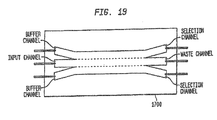
- An exemplary apparatus embodiment of the invention for separating a fluid mixture into constituent, non-motile components includes: a first sorting channel having a first inlet for a first flow and a second inlet for a second flow; the first sorting channel further having a first outlet for the first flow and a second outlet for the second flow, the first sorting channel further having means to maintain the first flow and second flow substantially non-turbulent, the first sorting channel adapted to allow a first component in the first flow, of a plurality of components in the first flow, to sediment into the second flow to form an enriched second flow and a depleted first flow, while concurrently maintaining a second component of the plurality of components in the first flow; a second, optically transparent sorting channel having a first optical inlet coupled to the first outlet for the first flow and having a first optical outlet, the second, optically transparent sorting channel further having a second optical inlet for a third flow and a second optical outlet for the third flow; and a holographic optical trap coupled to the second, optical
- Another apparatus or system for separating a plurality of components in a fluid comprises: an optically transparent sorting channel having a first inlet for a first flow and a second inlet for a second flow, the optically transparent sorting channel further having a first outlet for the first flow and a second outlet for the second flow; and a holographic optical trap system coupled to the optically transparent sorting channel, the holographic optical trap system adapted to generate a holographic optical trap to select and move a first component in the first flow, of a plurality of components in the first flow, into the second flow to form an enriched second flow and a depleted first flow, while a second component of the plurality of components is concurrently maintained in the first flow.
- Another method embodiment provides for separating a plurality of cells, comprising: providing a first flow having the plurality of cells; providing a second flow; contacting the first flow with the second flow to provide a first separation region; and differentially sedimenting a first cell of the plurality of cells into the second flow while concurrently maintaining a second cell of the plurality of cells in the first flow.
- the method generally also includes differentially removing the second flow having the first cell from the first flow having the second cell.
- the method may also provide for providing a third flow; contacting the first flow with the third flow to provide a second separation region; and differentially sedimenting the second cell of the plurality of cells into the third flow while concurrently maintaining a third cell of the plurality of cells in the first flow.
- a plurality of second cells may be holographically separated from the first flow, and a plurality of contaminants or biological debris may be holographically removed from the first flow.
- optical trapping which is a technology which has been used as a tool for manipulating microscopic objects.
- An accepted description of the effect is that tightly focused light, such as light focused by a high numerical aperture microscope lens, has a steep intensity gradient.
- Optical traps use the gradient forces of a beam of light to trap a particles based on its dielectric constant To minimize its energy, a particle having a dielectric constant higher than the surrounding medium will move to the region of an optical trap where the electric field is the highest.
- Optical trapping of the present invention is used to address cell sorting and purification (e . g ., from contaminants such as viruses and bacteria) in several ways.
- the forces exerted by optical traps on a material are sensitive to the exact distribution of the dielectric constant in that material - the optical force therefore depends on the composition and shape of the object.
- a diffractive optical element i.e., a phase shifting hologram utilizing either a transmission or a reflection geometry
- DOE diffractive optical element
- the wavefront is altered so that the downstream laser beam essentially becomes a large number of individual laser beams with relative positions and directions of travel fixed by the exact nature of the diffractive optical element.
- the present invention provides optical trapping by focusing a laser beam with a lens to create an optical trap wherein the lens has a numerical aperture less than 0.9, and preferably decreases until it is most preferably less than 0.1.
- Sorting using holographic laser steering involves establishing classes of identification for objects to be sorted, introducing an object to be sorted into a sorting area, and manipulating the object with a steered laser according to its identity class.
- the manipulation may be holding, moving, rotating, tagging or damaging the object in a way which differs based upon its identity class.
- the present invention provides a way of implementing a parallel approach to blood cell sorting and sperm cell sorting using holographic optical trapping.
- spectroscopy of a sample of biological material may be accomplished with an imaging illumination source suitable for either inelastic spectroscopy or polarized light back scattering, the former being useful for assessing chemical identity, and the latter being suited for measuring dimensions of internal structures such as the nucleus size.
- an imaging illumination source suitable for either inelastic spectroscopy or polarized light back scattering, the former being useful for assessing chemical identity, and the latter being suited for measuring dimensions of internal structures such as the nucleus size.
- cells are interrogated. The spectrum of those cells which had positive results ( i . e ., those cells which reacted with or bonded with a label) may be obtained by using this imaging illumination.
- a computer program may analyze the spectral data to identify the desired targets (i.e., cells bearing either an X or Y chromosome, or a suspected cancerous, pre-cancerous and/or non-cancerous cell types, etc.), then may apply the information to direct the phase patterning optical element (i.e., optical traps) to segregate or contain those desired or selected targets (i.e., cell types).
- the contained cells may be identified based on the reaction or binding of the contained cells with chemicals, or by using the natural fluorescence of the object, or the fluorescence of a substance associated with the object, as an identity tag or background tag.
- selection may be made, via computer and/or operator, of which cells to discard and which to collect.
- Manipulation of cells in general is made safer by having multiple beams available. Like a bed of nails, multiple tweezers ensure that less power is introduced at any particular spot in the cell. This eliminates hot spots and reduces the risk of damage. Any destructive two-photon processes benefit greatly since the absorption is proportional to the square of the laser power. Just adding a second tweezer decreases two-photon absorption in a particular spot by a factor of four. Trapping large cells involves a large amount of laser power for effective trapping. Putting the power into a single trap may cause immediate damage to the cell.
- a single cell may be manipulated by a line of tweezers, which lift the cell along the perimeter on one side. The resulting rotation allows a 360 degree view of the cell.
- a line of tweezers which lift the cell along the perimeter on one side. The resulting rotation allows a 360 degree view of the cell.
- Sorting with a wide field of view has many advantages such as higher throughput.
- standard tweezing in a WFOV wide field of view
- Tweezing with a wide field of view using holographic optical trapping may permit the ability to form exotic modes of light which greatly reduce the radiation pressure of the light beam.
- Vortex traps for example, have a dark center because the varying phases of light cancel in the center of the trap. This dark center means most of the rays of light which travel down the center of the beam no longer exist. It is exactly these beams which harbor most of the radiation pressure of the light, so their removal greatly mitigates the difficulty in axial trapping.
- Other modes e.g., donut modes, have the same advantage.
- the method and system lends itself to a semi-automated or automated process for tracking the movement and contents of each optical trap.
- movement may be monitored via an optical data stream which can be viewed, or converted to a video signal, monitored, or analyzed by visual inspection of an operator, spectroscopically, and/or by video monitoring.
- the optical data stream may also be processed by a photodectector to monitor intensity, or any suitable device to convert the optical data stream to a digital data stream adapted for use by a computer and program.
- the computer program controls the selection of cells and the generation of optical traps.
- the movement of cells is tracked based on predetermined movement of each optical trap caused by encoding the phase patterning optical element. Additionally, in some embodiments, a computer program maintains a record of each cell contained in each optical trap.
- the various embodiments of the present invention provide for separating components in a mixture, such as separating the various blood components of whole blood into corresponding fractions, such as a platelet fraction, a red blood cell fraction, a white blood cell fraction, and a plasma fraction.
- the various embodiments utilize one or more sorting channels, having a plurality of substantially laminar flows, allowing one or more components to differentially sediment from one flow into another, thereby separating the components into corresponding flows.
- the various components may be sorted further using optical mechanisms, such as holographic optical trapping.
- the various embodiments of the present invention thereby provide separation of components on a continuous basis, such as within a continuous, closed system, without the potential damage and contamination of prior art methods, particularly for fractionation of blood components.
- the continuous process of the present invention also provides significant time savings for blood fractionation.
- the present invention is also suitable for other cell sorting applications, such as separations of cancer cells from normal or healthy cells in, for example, bone marrow extractions.
- cell sorting applications such as separations of cancer cells from normal or healthy cells in, for example, bone marrow extractions.
- the various embodiments of the present invention have further applicability to other biological or medical areas, such as for separations of cells, sperm, viruses, bacteria, cellular organelles or subparts, globular structures, colloidal suspensions, lipids and lipid globules, gels, immiscible particles, blastomeres, aggregations of cells, microorganisms, and other biological materials.
- the component separation in accordance with the present invention may include cell "washing", in which contaminants (such as bacteria) are removed from cellular suspensions, which may be particularly useful in medical and food industry applications.
- cell "washing" in which contaminants (such as bacteria) are removed from cellular suspensions, which may be particularly useful in medical and food industry applications.
- prior art flow-based techniques have not recognized any applicability to sorting or separation of non-motile cellular components using variable sedimentation rates and optical manipulation.
- the apparatus, methods and systems of the present invention may be extended to other types of particulate, biological or cellular matter, which are capable of sedimenting or creaming within a fluid flow, or which are capable of being manipulated optically between different fluid flows.
- the methodology of the present invention could be utilized to separate non-motile or non-viable sperm cells from viable cells, by allowing the non-motile cells to sediment from a first flow into a second flow while retaining the motile cells in the first flow or allowing the motile cells to move to a third flow.
- cell separation may also be performed, such as separating islet cells from other types of pancreatic cells, or otherwise separating islet cell clusters of different sizes, through either or both flow separation or optical tweezing (trapping). Viruses, proteins and other large molecules having different sedimentation rates may also be separated with the present invention.
- the holographic optical trapping utilized with the various separation stages may also be particularly useful in these other types of cell or particle separations.
- the present invention has other medical applications as well.
- the various laminar flows discussed below may be utilized as part of a kidney dialysis process, in which whole blood is cleansed of waste products and returned to the patient.
- the present invention may be utilized for separations based upon diffusion, motility, and other types of gradients.
- the present invention may be utilized to move a species from one solution to another solution where separation by filtering or centrifugation is not practical or desirable.
- additional applications include isolating colloids of a given size from colloids of other sizes (for research or commercial applications), and washing particles such as cells, egg cells, etc. (effectively replacing the medium in which they are contained and removing contaminants), or washing particles such as nanotubes from a solution of salts and surfactants with a different salt concentration or without surfactants, for example.
- the action of separating species may rely on a number of physical properties of the objects including self-motility, self-diffusivity, free-fall velocity, or action under an external force, such as an electromagnetic field or a holographic optical trap.
- the properties which may be sorted upon include cell motility, cell viability, object size, object mass, object density, the tendency of particles to attract or repel one another or other objects in the flow, object charge, object surface chemistry, and the tendency of certain molecules to adhere to the object.
- FIG 1 is an illustration of a lateral view of an apparatus 100 in accordance with the present invention.
- the sorting apparatus 100 includes a sorting channel 110, a plurality of inlets 120 and a plurality of outlets 130.
- a corresponding fluid flow such as illustrated flows W, X, Y and Z, enters one of the inlets 120 and flows, substantially non-turbulently or otherwise as a laminar flow, across the sorting channel (or sorting region) 110, and out through a corresponding outlet 130, as illustrated.
- the apparatus 100 may be constructed of a plurality of materials, integrally or as discrete components, using a wide variety of materials, such as metals, ceramics, glass, and plastics.
- materials and fabrication methods are discussed in the third and fourth related applications, and include, for example, use of various polymers which cure under UV exposure.
- Other details are also provided in the third and fourth related applications, such as the use and selection of different types of pumps, such as syringe pumping, peristaltic pumping, gravity-driven pumping, and various combinations of pumping actions.
- the apparatus 100 when coupled with holographic trapping or other form of optical tweezing, the apparatus 100 (or 200) is transparent to the selected wavelength of the holographic generator, such as optically transparent when the holographic generator utilizes visible wavelengths.
- the apparatus 100 (200) should also be sterile and may also have a controlled temperature.
- the various fluid flows may be fed into the inlets 120 through a wide variety of means known to those of skill in the art and are within the scope of the present invention, including use of peristaltic pumps or a gravity feed, for example, and such means may also be utilized to control the flow rates of the various flows W, X, Y and Z.
- peristaltic pumps When peristaltic pumps are utilized, to maintain a constant flow rate and pressure, bubble-traps may be incorporated at the inlets 120 of the apparatus 100 (or 200).
- the various fluids utilized in the separation flows may be diluted or concentrated, to increase or decrease the volume of one of the solutions, or to impact the concentration of some dissolved or suspended material, or to impact physical properties of the solution such as its viscocity, temperature, or density.
- Examples for the apparatus 100, when used for blood sorting include: (a) dilution of the blood to reduce clogging or hydrodynamic interaction between blood cells, (b) extension of the volume of the blood or a blood fraction, (c) modification of the density of the blood, a blood fraction, or another solution which impacts the flow properties or separation behavior, (d) extension of the volume of a solution to maintain in increase fluid volume, especially in circumstances when fluid volume is being removed from the system.
- various chemical attractants and repellants may be added to the fluids, which may also be at different temperatures and viscosity levels, to improve sperm sorting.
- the various fluids utilized in the separation flows also may be "activated", such that some process is activated within the solution by some external influence or mixing with an external solution.
- external influences include: (a) applying an electric field, (b) applying a magnetic field, (c) exposing to light, (d) modifying the temperature, (e) introducing a chemical, (f) introducing a biological material, (g) shearing the solution, and (h) vibrating the solution.
- Examples of the activation which is caused by the external solution include: (a) alignment of particles, molecules, or cells, (b) polarization of one or more components of the solution, (c) cross-linking, (d) initiation or termination of chemical reaction, (e) initiation or termination of a biological response, (f) changing the type or rate of a chemical, physical, or biological response, or (g) causing a response or separation which depends upon the character of the particular component which is responding.
- Examples for the apparatus 100 when used for blood sorting, include: (a) addition of an agent to reduce clotting; (b) addition of agents to preserve viability or health of the blood solution or its components; (c) addition of agents which may augment the sorting process, such as by binding or collecting near certain components, thereby influencing one or more of their physical properties, including the addition of beads or other particles or polymers which may adhere to one or more species, and also including the introduction of salts or other materials which may influence the electrostatic interaction of materials or the hydrodynamic size or character of the materials; (d) addition of agents which may influence the flow properties, such as by changing the density, viscosity, surface tension, or other parameters; (e) addition of agents to enhance or suppress the aggregation of certain materials; and (f) addition of agents to enhance or suppress the adherence of certain materials to other materials or parts of the flow device.
- one of the fluid flows contains a plurality of components A, B, C and E.
- these components may be red blood cells (“RBC”), white blood cells (“WBC”), platelets, cellular debris and contaminants, all in plasma.
- RBC red blood cells
- WBC white blood cells
- platelets cellular debris and contaminants, all in plasma.
- many of the plurality of components have different sedimentation rates, typically measured using a Svedberg coefficient.
- RBCs have a comparatively greater sedimentation rate than platelets, and will be expected to sediment faster on a passive basis, such as due to gravitational or buoyant forces, without the intervention of other, active mechanisms, such as centrifugation.
- the plurality of components (such as cells or other particles) will sediment, moving from one flow to another.
- component A having the comparatively greatest sedimentation rate is illustrated as having moved from flow W to the lowest flow Z
- component B having the comparatively next highest sedimentation rate is illustrated as having moved from flow W to the flow Y (above Z)
- component C having a comparatively smaller sedimentation rate is illustrated as having moved from flow W to the flow X (above Y)
- component E having the comparatively smallest sedimentation rate is illustrated as having remained in flow W (above Y).
- each of these components may be separated into a corresponding flow, and isolated from each other as each flow exits through its corresponding outlet 130.
- that flow is differentially removed from the other flows, i.e., the flow is removed while the other flow remains intact or is otherwise separately removed from the remaining flows.
- this differential removal may be concurrent, namely, all flows removed concurrently or continuously.
- the various blood fractions may be separated from each other, with red blood cells sedimenting fastest and represented by component A (e . g ., 4.59 ⁇ m/s), white blood cells sedimenting at a slightly lower rate and represented by component B ( e . g ., 2.28 ⁇ m/s), platelets sedimenting at a comparatively slower rate and represented by component C ( e . g ., 0.055 ⁇ m/s), and plasma continuing to comprise flow W and represented by component E.
- Each blood fraction may then be removed through a corresponding outlet 130.
- the sorting channel (or sorting region) 110 of apparatus 11 has a varied length "L” parallel to the direction of flow, a depth “D” perpendicular to the direction of flow, and a width "WW", illustrated as extending into the page (and designated WW to avoid confusion with the W flow). These various dimensions may be selected based on a plurality of factors, particularly the flow rates and the sedimentation rates of the components of interest.
- the total length of the sorting channel should be long enough to differentially remove the component having the comparatively slowest sedimentation rate, illustrated as component C in Figure 2 , with shorter lengths corresponding to other flows for separation of components having faster sedimentation rates, as illustrated for flow Z having component A and flow Y having component B.
- the present invention is further distinguished from the prior art by having considerably more latitude or tolerance for aspect ratios, while nonetheless maintaining a substantially laminar flow.
- the aspect ratio of length to width may vary from about 5 (or more) to 1 (5:1), with the length being greater than the width, to about 1 (or less) to 2 (the length being smaller than the width).
- a preferred length to width aspect ratio is about 2: l, and may vary from 3:1 to 1:2.
- Flow rates may also vary between the plurality of flows utilized in apparatus 100. For example, higher flow rates in the lower level flows (such as Y and Z) may tend to compress the flows W and X, resulting in a shorter distance that certain components must traverse to sediment into the flows Y and Z.
- the sedimentation of components through the various flows of the apparatus 100 is typically rate zonal, that is, based upon both relative density and size of the components to be separated, as well as the material's shape and electrostatic properties. Under other conditions, however, such as slower flow rates, thinner flow depths, and/or longer sorting channels 100, the sedimentation may also be isopycnic, that is, based only upon relative density of the components.
- the various fluids comprising the flows W, X, Y and Z may be selected and adjusted to create desired density gradients to match the component densities for the selected separations.
- the various fluids comprising the flows W, X, Y and Z may be selected and adjusted to each have a different, increasing or decreasing density, creating a stepped density gradient, with various particles sedimenting to the appropriate step.
- the density gradient will effectively become continuous, with a corresponding ability for fine-grained separation.
- the various fluids comprising the generally laminar flows may also be selected based on suitable criteria for the particular desired component separation.
- the various flows may be comprised of whole blood, such as for flow W, and plasma or buffering solutions for the remaining flows.
- the various fluids may also be preprocessed prior to entry through the inlets 120, such as through dilution, addition of other components such as additives (such as anticoagulants, flocculants, or binding agents), viscosity or other flow property manipulation, or preprocessed through other separation techniques.
- whole blood may be preprocessed to initially remove some red blood cells or to add an anticoagulant such as sodium citrate.
- the apparatus 100 may be implemented with any number of flows and corresponding fluid inlets 120 and outlets 130.
- One limitation to the number of fluid flows is based on the ability to maintain each flow in a substantially laminar or non-turbulent manner, such that each interface between flows is substantially non-turbulent, to minimize any unwanted mixing of flows.
- there also may be relative density considerations for the fluids comprising the flows which could also result in limiting the number of flows utilized in a given stage of separation.
- the various apparatus 100 may be further coupled to additional apparatus 100 (200, below), in parallel for higher throughput, and in series for additional separation stages, such as for increased purity levels.
- the various apparatus 100 may also be combined with non-sedimentation separations, or be coupled in series with subsequent separations using non-sedimentation mechanisms, with additional separation of components between flows accomplished, for example, using optical forces such as holographic optical trapping of the fifth related application, incorporated herein by reference.
- These various apparatus 100, 200 or 300 may have different dimensions and different numbers of channels or flows.
- Figure 2 (and also Figure 13 ) is a general illustration of using such optical forces created by holographic or optical trapping for additional component separation in an apparatus 200. Creation and manipulation of the plurality of holographic optical traps 210 is explained in greater detail below with reference to Figures 4 and S, with apparatus 200 forming the sample 506 of Figure 5 .
- Two flows W and X are illustrated in Figure 2 , with flow W initially having two components A and B.
- Holographic optical traps 210 (illustrated as conic sections in Figure 2 ) are then utilized to capture "A" components, and move them into flow X.
- Such optical trapping is particularly useful for increased purification of a particular fraction, particularly for fractions having insufficient differentiation based on sedimentation rates.
- optical trapping is also particularly useful for removal of undesirable components, such as cellular debris and other impurities.
- the optical trapping may be particularly accurate in removing undesired components. For example, a comparatively small portion of white blood cells may not have sedimented fast enough, resulting in some white blood cell contamination of a platelet fraction.
- Optical trapping may be utilized to select and move the white blood cells into a separate flow, increasing the purity of the platelet fraction.
- platelets and RBC optically manipulate (or "tweeze") better than white blood cells.
- Using lower numerical apertures in the system 500 discussed below, however, significantly improves optical manipulation of white blood cells.
- the apparatus 100, 200 or 300 When implemented in conjunction with optical traps, the apparatus 100, 200 or 300 should be embodied utilizing an optically transparent material, for the selected optical wavelength. When the holographic traps are implemented at other wavelengths, other correspondingly transparent materials may be utilized which are suitable for the selected wavelength.
- the apparatus 100, 200 or 300 is then implemented and placed in the location of the sample 506 illustrated in Figure 5 , with the system 500 utilized to perform the holographic optical trapping as one of or as part of a separation stage of the present invention.
- FIG. 3 is a diagram illustrating a closed, two-stage system 300 for blood or other component separation in accordance with the present invention.
- a first stage 305 blood components from a selected donor flow through inlet 315 to form a first flow, and plasma is returned (or primed on initial start up) through inlet 320 to form a second flow.
- the first and second flows are non-turbulent and otherwise laminar flows, and make non-turbulent contact with each other in first separation region 325, forming a non-turbulent interface region between the two flows.
- both red blood cells and white blood cells sediment from the first flow into the second flow, and are collected in reservoir 330 for other uses (such as medical uses for packed cells) or for return to the selected donor.
- both the length of the first separation region 325 and the flow rate of the first flow are predetermined such that both red blood cells and white blood cells have sufficient time to passively sediment into the second flow, under gravitational and buoyant forces.
- the first flow now substantially depleted of both red blood cells and white blood cells, flows non-turbulently on a continuous path into a second separation stage 310.
- the first flow enters a second separation region 340 with a third flow from inlet 335.
- the third flow is also comprised of plasma from the selected donor in the exemplary embodiment.
- the platelets remaining in flow one passively sediment into flow three, and are collected with the plasma of flow three in reservoir 345 for medical use, for example.
- the further depleted flow one is then recirculated from outlet 350 back to inlets 320 and 335, to form the first and third flows, respectively.
- the system 300 may be primed with donor plasma at system start-up by, for example, centrifuging a portion of the selected donor's blood, or by initially using another biocompatible, non-toxic liquid until a depleted flow one (substantially or predominantly plasma) is generated at outlet 350.
- an additional holographic trapping separation stage may also be utilized to aid in the separation of these blood fractions.
- a holographic trapping separation stage may be utilized in lieu of, or in addition to, second stage 310.
- flow two may be subjected to a second (or more) stage separation, in addition to or in lieu of the additional separation stage of flow one.
- additional separation stages may be utilized in series or in parallel.
- separation regions are regions where materials are partially or fully sorted or separated based upon some material property.
- the separation regions may employ one or more of the following techniques, individually, serially, or simultaneously, in any of the various flows in the embodiments of the invention:
- exemplary separations for blood sorting include: (a) separation of one or more blood cell types from some or all of the other blood cell types and/or from the blood plasma or fluid medium by sedimentation rate zonal separation; (b) separation as in (a) but with isopycnic separation; (c) removal of just the red blood cells (RBCs) or the RBCs and white blood cells (WBCs) from the solution by sedimentation; (d) concentration of the platelets from the plasma using sedimentation; (e) extraction of the RBCs using dielectrophoresis, electrophoresis, or magnetic separation; (f) concentration or extraction of platelets from a solution using optical techniques including optical tweezers and optical fractionation; and (g) separation of blood components using agents which may bind to a particular cell type (such as functionalized beads) and be acted upon by any of the above separation techniques, after which the agent may or may not be unbound from the cell type.
- RBCs red blood cells
- WBCs white blood cells
- a combined approach may be the most effective, such as: first extract most of the RBCs and WBCs using sedimentation rate zonal separation, then extraction and concentration of the platelets using optical fractionation, discussed below.
- the optical fractionation step will act not only to concentrate the platelets (which could also, for example, be done by a centrifugation step at the end), but will provide a second step which will exert strong suppression on the WBCs accidentally collected with the platelets, for the example of platelet aphoresis.
- concentration steps may also include filtering, such as to filter WBC from a platelet fraction or a plasma fraction.
- apparatus 300 has been described with respect to blood fractionation, it will be understood by those of skill in the art that the apparatus 300 may be utilized for a wide variety of separations, in addition to such blood fractionation. In addition, apparatus 300 may also be considered one particular embodiment of series-connected separation stages of the present invention.
- Cell “washing” is also a significant application of the apparatus 100, 200 or 300. Such washing may include a change of media, for storage, preservation, or other medical purposes. Such washing may also consist of removing a media containing contaminants such as bacteria, by separating the cells of interest into another media flow free of such contamination. As indicated above, sperm separation is also a significant application of the apparatus 100, 200 or 300.
- Portions of, or outputs from, the sorting device 100, 200 or 300 may be inspected optically. This may be direct visual imaging, such as with a camera, utilizing direct bright-light imaging or fluorescent imaging. Or, it may be more sophisticated techniques such as spectroscopy, transmission spectroscopy, spectral imaging, or scattering such as dynamic light scattering or diffusive wave spectroscopy. In many cases, these inspection regions may be incorporated directly into the flow device to characterize the inputs, outputs, or intermediate steps. They may be for diagnostics or record-keeping, or they may be used to inform the overall process, such as for feedback on how processing is done or on the speed of flow or amount of each solution to use.
- the optical inspection regions may be used in conjunction with additives, such as chemicals which bind to or affect parts of the solution or beads which are functionalized to bind and/or fluoresce in the presence of certain materials or diseases.
- additives such as chemicals which bind to or affect parts of the solution or beads which are functionalized to bind and/or fluoresce in the presence of certain materials or diseases.
- these techniques may be used to measure cell concentrations, to detect disease, or to detect other parameters which characterize the blood.
- Portions of, or outputs from, the sorting device 100, 200 or 300 also may be characterized electronically.
- a portion of the sorting device may have electronic devices embedded.
- Example electronic devices may include: (a) capacitors, (b) electronic flow meters, (c) resistance meters for determining the bulk conductivity of the fluid, from which concentrations or compositions may be measured, or (d) pH measuring devices.
- measurements of cell concentration, iron content, flow rates, total cell counts, electrolyte concentration, pH, and other parameters may be a valuable part of a sorting device.
- the flow components of the sorting device 100, 200 or 300 may be passive, being completely controlled externally by the flow rates of the inputs and outputs. Alternatively, there may be active flow components embedded in the device (not separately illustrated), such as valves which may be partially or fully opened or closed using electronic, optical, thermal, mechanical, or other influence.
- the sorting device 100, 200 or 300 may have an integrated method for storing and/or delivering one or more solutions.
- a consumable sorting device may be manufactured to have a deformable membrane on a side of a reservoir. This reservoir may be filled with a solution, such as a buffering agent which is biologically compatible with the patient and which may be used to dilute the blood.
- Another example is that it could be filled with an anti-coagulant.
- the delivery and/or use of one such fluid may be actively controlled by mechanical influence, pressing on the membrane to deliver the fluid.
- another mechanism may be used to deliver the fluid.
- the integration of various fluid solutions needed at differing steps in the sorting Integrating these components may result in substantial simplification and reduction of the total cost of ownership and operation. It may also reduce the risk of contamination and error.
- Such reservoirs holding input and buffering solutions or fluids are illustrated, for example, in Figures 14 and 15 .
- the device 100, 200 or 300 may include areas (not separately illustrated) where biological or chemical investigation of one or more of the fluids or fluid components. This may include measurements of pH, the presence of certain biological or chemical materials, or other measurements. For the application of blood sorting, this may include detection of disease, characterization of concentrations of various cell types or materials in the plasma, characterization of iron content, determination of blood type, or other evaluation of blood quality, type, or category.
- the device 100, 200 or 300 may include a region (not separately illustrated) which sterilizes the solution using optical methods, such as exposure to UV light, or other methods.
- the sterilization may act upon solutions which are initially part of the device or added to it for buffering, washing, diluting, or other impacts on the sample solution. Or, the sterilization may act upon part or all of the sample solution being processed.
- optical sterilization of the solutions used in the device other than whole blood may be important. Also, sterilization of the blood or certain fractions of the
- the device 100, 200 or 300 may be comprised of materials such that one or more surfaces have been constructed so as to interact physically or chemically with certain materials.
- a surface may be functionalized so that certain materials adhere to it, for the purpose of extracting these materials from the solution or for the purpose of diagnostics.
- functionalized surfaces may be used to extract unwanted materials from certain fractions. Alternately, they may be used to collect materials which are at low concentration for the purpose of measuring the degree to which a material or a type of material is present, such as for disease detection.
- the sorting device 100, 200 or 300 may contain regions where sorting acts in parallel but without physical walls to separate the flows.
- a parallel sorting region may have multiple inlets 120 and outlets 130, some of which are functionally similar to each other. Instead of having physical dividers distinguishing the multiple parallel sorters, the division occurs as a consequence of the physical properties of the solutions and the flow.
- the contacting parallelized sorters regions may yield high sorting rates with more simple and cheap devices. They may also act to avoid contact with surfaces. For blood sorting, these regions would be used to minimize contact with surfaces and to maximize sorting rate while reducing costs and complexity.
- the device 100, 200 or 300 may have regions which are designed to regulate flow rate or the flow character (not separately illustrated). For example, in many cases laminar flow is required, and often a particular flow profile is desired. In other cases, several regions of the device should have identical flow rates and behavior. For these reasons, areas with shapes and other properties to influence flow behavior are often needed. In some cases, this is done by making very symmetric flow designs. In other cases, large changes in the diameter of flow regions and/or the existence of reservoirs help to maintain uniform flow rates. In other cases, very carefully designed channels provide the exact balancing of flow rates needed. To maintain laminar flow, areas where slow changes in channel size occur may be important. Obstacles or dividers may also act to maintain laminar flow. For blood sorting, flow regulation is important to achieve high sorting rates while maintaining the yield and purity of the fractions.
- the device 100, 200 or 300 may contain regions which are designed for mechanical mixing of the fluids, such as regions which encourage turbulence.
- regions which encourage turbulence For example, a region with a fast narrow stream entering a region with a large dimension may produce turbulent flow and mixing.
- a mixing region may mix a diluent or anticoagulant with the blood, or may mix other solutions together as needed.
- the device 100, 200 or 300 may contain regions which align cells or materials in a certain way (not separately illustrated). This is sometimes done through shear flows, but may also be done by imposing external fields such as electric fields.
- the device 100, 200 or 300 may contain regions designed to lyse cells or break up materials (not separately illustrated). This may be done through shear flows, vibration, forcing through an orifice, electrical, or other means. For blood sorting, this may be valuable for the elimination of certain cell types or aggregates which may form. It may also be valuable for diagnostic purposes, such as disease detection or measurement of parameters which pertain to the contents of cells.
- the device may contain regions (not separately illustrated) which swell or dehydrate cells or objects, such as by introducing agents which change the osmotic pressure or by changing the physical pressure. This may be done, for example, as a step prior to isopycnic sorting to adjust the density of the cells or objects. It may also be done to kill or shock certain components. For the example of blood sorting, this may be done as a later stage purification step to remove or neutralize undesired components.
- the device 100, 200 or 300 may contain regions which heat or cool one or more solutions (not separately illustrated). This may be done for its impact on physical properties, such as viscosity or density. Or, it may be done for its impact on chemical properties, such as chemical reaction rates or chemical stability. Or, it may be done for its impact on biological properties, such as motility, metabolism rate, or viability. For example, one fluid flow may be at a higher temperature than another, causing motile sperm to move away from the hotter fluid to the cooler fluid. Also, it may be done for system-level compatibility, such as in preparation for the following processing step. For the example of blood sorting, solutions which are returned to the patient may be maintained at an appropriate temperature to avoid chilling the patient. Solutions which are to be stored may be cooled during processing to preserve those fractions or prepare them for the next processing or storage step.
- the device 100, 200 or 300 may contain reservoirs which serve to store solutions which will be used during the process run, or which may be generated during the process run (not separately illustrated). Having these reservoirs integral to the sorting device simplifies the use of the device and reduces the need for additional parts.
- reservoirs may contain anticoagulants, diluents, dilutants, and any other solutions needed in the process. Reservoirs may also be incorporated which will hold the sorted fractions or waste fractions.
- the device 100, 200 or 300 may contain regions which enhance mixing by diffusion (not separately illustrated). For example, when mixing by contacting two laminar flows, parallelizing into many narrow contacting flows enhances the overall mixing rate by diffusion. For the example of blood sorting, diffusive mixing regions may be used to mix diluent, anticoagulant, or other solutions with whole blood or blood fractions.
- the device may contain regions with bubble traps to remove air bubbles from the system. This may be done by having a region where air bubbles are able to rise from a region with flow to a region above the primary flow region. For the example of blood sorting, this may be done in a simple way to guarantee that small air bubbles from the loading or running of the system do not pass on to the patient or the collection samples.
- the device 100, 200 or 300 may contain regions which act to suppress any pulsation in the flow (not separately illustrated), such as that which occurs when peristaltic pumps are used.
- One way to suppress pulsation is to incorporate a "bubble-trap" into the device.
- the presence of an air pocket, which is in contact with the fluid, allows for compression of the air pocket as pressure in the fluid increases and decreases.
- the air pocket acts as a shock absorber, smoothing out the flow.
- Other devices may be used as well, such as a flexible membrane which may bend under higher pressures, thereby smoothing out the pressure and flow rate.
- pulsation reduction regions will yield more precise and smooth flows, and therefore higher sorting rates, purity, and yield.
- the device 100, 200 or 300 may contain regions which reveal the state of the device (not separately illustrated). For a consumable, it may indicate whether the device has been sterilized or whether it has been used. For the application of blood sorting, one would want indicators to confirm both that the sorting device has been sterilized and that the device has not yet been used or contaminated.
- the sorting device 100, 200 or 300, or overall sorting system may contain mechanisms for precise leveling of the flow sorter. This is important because for some sorters, buoyant forces may cause unintentional flows and have negative impacts on sorting yields and purity in cases where the device is not precisely leveled. Additionally, the sorting device may contain components which reveal whether it is well-balanced, or the degree to which it is balanced. For example, it may have an electronic or gravity-based balance incorporated in the sorting device itself.
- One example of such a device is a shaped channel with fluid and an air bubble in it. The position of the air bubble may reveal the angle of the tilt of the device. Another such device may use a metal ball in a track to reveal the tilt angle.
- Another manifestation is to use an optical alignment, such as bouncing a light source off a surface or passing a light source through a wedge to identify its orientation.
- leveling controls and indicators are significant to guarantee high-yield and high-purity products.
- the sorting device 100, 200 or 300, or overall sorting system may contain mechanisms for maintaining uniform and/or constant temperatures of the device and/or the solution (not separately illustrated). This is important to eliminate thermally-induced buoyant forces which may cause unintentional flows and have negative impacts on sorting yields and purity. Additionally, the sorting device may have indicators in it, or in the sorting system as a whole, which indicate the temperature and/or temperature uniformity of one or more components. For the application of blood sorting, temperature uniformity controls and indicators may be significant to guarantee high-yield and high-purity products.
- the sorting device 100, 200 or 300, or overall sorting system may contain mechanisms for measuring the level and concentration of one or more input or output solution (not separately illustrated). These measurements may be used to gauge the speed of operation, completion time, error state, for general monitoring, or for other applications. For the application of blood sorting, level and concentration indicators may be used to identify when sufficient sample has been collected or to detect when a failure or depletion of a solution has occurred.
- the sorting device 100, 200 or 300, or overall sorting system may have a method for priming the system with one or more fluids using standard bottom-up filling or evacuation. Purging may similarly be done by draining the device or by flowing a solution through it. At the early stages of a sorting run, the priming solution may be discarded until a time when the priming solution has been mostly exhausted and the desired solution is obtained. Similarly, at the late stages of a sorting run, a fluid may be used to push the sorted material through the system to minimize waste and maximize yield.
- FIG. 26 is a block diagram illustrating an exemplary sorting and separation system 2600 in accordance with one embodiment consistent with the present invention.
- This general purpose sorter would vary the flow rates on each input and output channel in response to user controls (user input 2610).
- the user controls would allow the user to determine the range of S values that leave through each output channel.
- This device would be a valuable lab tool for performing various purifications, washing, separations, and diagnostic evaluations of samples. It would also be an important platform for research, development, and prototyping applications.
- the sorting hardware may be contained within a single enclosure which may or may not be temperature controlled.
- This hardware consists of: (1) Consumable flow plate 2620 (e . g ., sorting device 100, 200 or 300) with several inputs and outputs, (2) several computer-controllable peristaltic pumps 2625, (3) temperature control and monitoring apparatus 2630, (4) reservoir holders (or reservoirs 2635 and 2640).
- the sorting is controlled by a computer 2650 which monitors and controls the flow rates, temperature, and any other additional components which may be included for controlling or diagnostics purposes.
- the flow plate 2620 may be a simple general-purpose device, suitable for many applications, as discussed above. It may have two to four inputs and two to ten outputs. It may be provided in a sterile, primed, and sealed state. The reservoirs may be part of the flow plate, or they may be separate sterile components which are attached to the flow plate before use. All or a portion of the central sorting region may be covered with a cover glass to allow manipulation with optical tweezers, optical dielectrophoresis, or laser killing/cutting of samples.
- the computer 2650 control does measurements and control of the hardware for pumping and temperature control. It may also interface other hardware components that may be added.
- the user software provides a very convenient front end to the simultaneous control of all the pumps. It would do the necessary math to control each pump rate to give the user the desired flows for the particular sorting application.
- This system 2600 would be general enough to allow for many sorting applications, either independently or in conjunction with an additional laser apparatus. These applications include but is not limited to: cell identification by fluorescence, cell killing by high intensity laser exposure, cell fractionation using optical dielectrophoresis, motile cell fractionation based on motility, passive sorting of objects by diffusivity, and passive fluidic zonal density sorting.
- the passive fluidic zonal density sorting applications which are numerous and extend over a broad range, can generally be done with minimal or no additional hardware.
- Exemplary uses and applications for such a general purpose sorting system 2600, using passive fluidic sorting and/or use of optical gradient forces are outlined in the following table: Category General Specific Washing - Removing Components Cell Washing for Disease Removal Removing debris, bacteria, and viruses from human sperm Removing debris, bacteria, and viruses from animal sperm Aggregate Removal Removing undesirable clumps of materials in industrial processes Removing aggregated colloids in solution Removing coarsened droplets in an emulsion Precursor Removal Removing precursor materials from a halted growth process Separating differentiated cells Removing blastocytes from incubating sperms and eggs Washing - Changing Media Cell Processing Changing osmotic conditions - Cell swelling or shrinking Cell staining Automated multi-step cell experiments or processing Sperm fluoridation to suppress activity Material Processing Bead dying Assays Object-solution response assays Diagnostics Characterizing Monodisperse Solutions Measuring S (Svedberg coefficient
- Sorting osteoclasts, osteoplasts, osteoblasts Sorting white blood cells as a treatment for white blood cell diseases (e.g., high WBC such as leukemia or lymphomia, or low WBC) Sorting sickle cells from normal red blood cells as a treatment for sickle-cell anemia Cell Cluster Sorting Sorting islet cell clusters by size (diabetes) Removing cell clusters from single cells Cell State Sorting Removing infected cells from healthy cells Removing living cells from dead cells Separating proliferating and non-proliferating cells Isolating viable sperm from inviable sperm Isolating Cells From Biopsies Purifying bone marrow cells from blood in marrow biopsy Colloids Fractionation Sorting colloids by size, density, composition, S, etc. Sorting Variants of One Cell Type Sorting multizygotic sperm from normal sperm Removing most severe RBCs in sickle cell anemia patients
- FIG. 27 is a block diagram illustrating an exemplary bioreactor product purification and separation system 2700 in accordance with one embodiment consistent with the present invention.
- a bioreactor is used in the production of monoclonal antibodies, recombinant protein products, viruses and viral antigens, and viable cell mass. The most common use for a bioreactor is in the production of various drug therapies.
- a bioreactor basically works by allowing cells to grow in high concentrations in ideal conditions. While the cells are growing a culture medium is flowed through the bioreactor. This medium collects the cell's waste products, as well as provides nutrition for the cells. The medium that has been flowed through the bioreactor is then processed for the product.
- the waste filled medium is filtered through many different processes, one of which is through bead column chromatography. This process takes a long time to complete, is very expensive, and does not yield a large percentage of the final product.
- the present invention solves all three of these problems.
- the bioreactor product purifier 2700 would quickly, easily, and accurately remove the wanted product from the waste medium.
- the waste media would be flowed into the sorter through one channel 2701
- a solution of beads coated with the proper affinity sites would be flowed into the sorter through the second channel 2702.
- Both channels would lead to a first mixer 2705 in which they would be mixed together.
- the product would bind to the sites on the beads.
- the mixture would then flow through a first separation region 2710 along with an input buffer (input channel 2715).
- the beads with selected product output through channel 2718 and the waste solution would be output through channel 2716 and discarded.
- Passive diffusive sorting succeeds because the large beads stay in one channel while the lighter molecules diffuse to the other side of the channel.
- the bead solution is then be flowed through channel 2718 as a denaturing solution is flowed through a second channel 2720. Both of these channels would flow to a second mixer 2725 where they would be mixed together. While they were being mixed, the denaturing solution would break the bond between the product and the beads.
- the solution After mixing, the solution would be flowed through an output channel 2735 along with another buffer solution through channel 2730, into second separation region 2740.
- the beads would be flowed through the bottom channel 2745 and be discarded as waste.
- the purified and recovered product would be flowed through the top channel 2750. This method of purification should reduce costs, reduce time, and increase the yield of final product.
- FIG. 4 schematically illustrates a holographic optical trapping system 400, generally used in conjunction with an apparatus 100, 200 or 300, according to one embodiment consistent with the present invention. Additional detail concerning holographic optical trapping is available in the fifth related application.
- a holographic optical trapping apparatus or system 400 as illustrated in Figure 4 light is incident from a laser system, and enters as shown by the downward arrow, to power the system 400.
- a phase patterning optical element 401 is preferably a dynamic optical element (DOE), with a dynamic surface, which is also a phase-only spatial light modulator (SLM) such as the "PAL-SLM series X7665,” manufactured by Hamamatsu of Japan, the "SLM 512SA7” or the “SLM 512SA15” both manufactured by Boulder Nonlinear Systems of Boulder, Colorado.
- SLM phase-only spatial light modulator
- These dynamic phase patterned optical elements 401 are computer-controlled to generate beamlets by a hologram encoded in the medium which may be varied to generate the beamlets and select the form of the beamlets.
- a phase pattern 402 generated on the lower left of Figure 4 produces the traps 403 shown in the lower right filled with 1 ⁇ m diameter silica spheres 404 suspended in water 405.
- the system 400 is controlled by the dynamic hologram shown below on the left.
- the laser beam travels through lenses 406, 407, to dichroic mirror 408.
- the beam splitter 408 is constructed of a dichroic mirror, a photonic band gap mirror, omni directional mirror, or other similar device.
- the beam splitter 408 selectively reflects the wavelength of light used to form the optical traps 403 and transmits other wavelengths.
- the portion of light reflected from the area of the beam splitter 408 is then passed through an area of an encoded phase patterning optical element disposed substantially in a plane conjugate to a planar back aperture of a focusing (objective) lens 409.
- single beam optical trapping also called laser or optical tweezers
- a high numerical aperture lens was necessary for acceptable optical traps.
- a basis for this thinking was that, for optical trapping, one uses the gradient in the electric field of the impinging light to trap the particle. In order to have a large trapping force it has been thought necessary to have a large gradient in the electric field (or number density of rays). The way that one usually accomplishes this is to pass the light field through a high numerical aperture lens.
- the invention of the fifth related application provides a low numerical aperture lens as, for example, the objective lens 409 in Figure 4 .
- the ability to observe and trap in this situation could be useful in any application where one would benefit from a large field of view given by a low magnification lens, such as placing microscopic manufactured parts or working with large numbers of objects, such as cells, for example.
- 3 micron silica spheres 104 suspended in water 105 were trapped with lenses 109 with an unprecedented low numerical aperture.
- the lenses 109 used were manufactured by Nikon: (a) Plan 4x with an NA of 0.10 ; and (b) Plan 10x with an NA of 0.25.
- Suitable phase patterning optical elements are characterized as transmissive or reflective depending on how they direct the focused beam of light or other source of energy.
- Transmissive diffractive optical elements transmit the beam of light or other source of energy, while reflective diffractive optical elements reflect the beam.
- the phase patterning optical element 401 may also be categorized as having a static or a dynamic surface.
- suitable static phase patterning optical elements include those with one or more fixed surface regions, such as gratings, including diffraction gratings, reflective gratings, and transmissive gratings, holograms, including polychromatic holograms, stencils, light shaping holographic filters, polychromatic holograms, lenses, mirrors, prisms, waveplates and the like.
- the static, transmissive phase patterning optical element is characterized by a fixed surface.
- the phase patterning optical element 401 itself is movable, thereby allowing for the selection of one more of the fixed surface regions by moving the phase patterning optical element 401 relative to the laser beam to select the appropriate region.
- the static phase patterning optical element may be attached to a spindle and rotated with a controlled electric motor (not shown).
- the static phase patterning optical element has a fixed surface and discrete regions.
- the fixed surface has a nonhomogeneous surface containing substantially continuously varying regions, or a combination of discrete regions, and substantially continuously varying regions.
- suitable dynamic phase patterning optical elements having a time dependent aspect to their function include computer-generated diffractive patterns, phase-shifting materials, liquid crystal phase-shifting arrays, micro-mirror arrays, including piston mode micro-mirror arrays, spatial light modulators, electro-optic deflectors, accousto-optic modulators, deformable mirrors, reflective MEMS arrays and the like.
- the medium 405 which comprises the phase patterning optical element 401 encodes a hologram which may be altered, to impart a patterned phase shift to the focused beam of light which results in a corresponding change in the phase profile of the focused beam of light, such as diffraction, or convergence. Additionally, the medium 405 may be altered to produce a change in the location of the optical traps 403. It is an advantage of dynamic phase patterning optical elements 401, that the medium 405 may be altered to independently move each optical trap 403.
- overfilling the back aperture by less than about 15 percent is useful to form optical traps with greater intensity at the periphery, than optical traps formed without overfilling the back aperture.
- the form of an optical trap may be changed from its original form to that of a point optical trap, an optical vortex, Bessel beam, an optical bottle, an optical rotator or a light cage
- the optical trap may be moved in two or three dimensions.
- the phase patterning optical element is also useful to impart a particular topological mode to the laser light, for example, by converting a Gaussian into a Gauss-Laguerre mode. Accordingly, one beamlet may be formed into a Gauss-Laguerre mode while another beamlet may be formed in a Gaussian mode.
- Gauss-Laguerre modes greatly enhances trapping by reducing radiation pressure.
- the current instrument design uses a high resolution CCD camera for the primary imaging system 110.
- the main advantage of the CCD camera (see reference numeral 511 in Figure 5 ) is the favorable cost/performance ratio since this technology is a mature one.
- Another advantage of CCD cameras is their wide dynamic range and the ease of generating digital output.
- the images are viewed on a computer screen (see reference numeral 510 in Figure 5 ) to provide both a frame of reference for selecting the location of the traps as well as to minimize the possibility of inadvertent exposure of the operator to the laser.
- the user interface consists of a computer screen which displays the field of view acquired by the CCD camera.
- the user designates the loci of the traps with a mouse. There is also an option to delete a location.
- the user is also able to specify the power per trap so as to be able to avoid specimen damage.
- the purpose of designating the loci of the traps is to provide input for the hologram calculation.
- the hologram is essentially a function whose Fourier transform produces the desired trap array.
- this function is a phase object (i.e., an object that changes the phase of the wavefront without absorbing any energy).
- the present invention also includes the capability of changing the height of the traps. If a laser beam is parallel to the optical axis of the objective lens 409, then a trap forms at the same height as the focal plane of the lens 409. Changing the height of a trap is accomplished by adjusting the hologram so that the beam of light forming a trap is slightly converging (or diverging) as it enters the objective lens 409 of the microscope. Adjusting the height of a trap is possible using lenses but only a holographic optical trapping (HOT) allows the height of each individual trap to be adjusted independently of any other trap. This is accomplished by the computer program adjusting the phase modulation caused by the liquid crystal hologram.
- HAT holographic optical trapping
- the sample chamber 700 (see FIGS. 7A and 7B ) of the present invention is inexpensive and disposable. Although the sample chamber 700 of the present invention is described below, another object of the present invention is to create a flexible design that may be changed for differing applications. In addition to the sample chamber 700, the various other separation stages of the invention may be utilized, such as an apparatus 100, 200 or 300, or the other separation stages discussed below with reference to Figures 13- 24 and 26-27 .
- the sample chamber 700 lies on the surface of a microscope slide 701.
- the sample chamber 700 contains a series of channels 703 for introducing specimens or objects.
- the channels 703 are connected to supply and collection reservoirs by thin tubing 704 (commercially available). Samples or objects will be suspended in a liquid medium and will be introduced into the working area via the channels 703.
- the sample chamber 700 is covered by a cover slip 705.
- a poly(dimethyl siloxane) (PDMS) resin is used to fabricate the chamber 700.
- the process involves creating the desired pattern of channels 703 on a computer using standard CAD/CAM methods and transferring the pattern to a photomask using conventional photoresist/etching techniques.
- the photomask is then used as a negative mask to create an inverse pattern of channels which are etched on a silicon wafer.
- the depth of the channels 703 is controlled by the etch time.
- the silicon wafer is a negative replica of the actual sample chamber 700.
- the final step consists of creating the positive sample chamber 700 by pouring PDMS onto the wafer and polymerizing. This results in a PDMS mold which is bonded to a glass slide 701 and overlaid with a cover slip 705.
- the glass to PDMA bonding is effected with an oxygen etch which activates the exposed surfaces.
- the PDMS solution/hardner is maintained under a vacuum in order to prevent bubble formation.
- the silicon wafer is silanized to prevent the PDMS from sticking to the wafer.
- steps involving cleaning the replicas and maintaining proper environmental controls These represent standard technology.
- the channels 703 are connected to microbore tubing 704 using small syringe needles 706 held using glue 714, which are inserted through the PDMS mold into small circular wells 707 which connect to each channel 703. Sample solutions are introduced into the channel 703 using micropumps 708.
- Figure 7B shows a diagram of a typical arrangement for the introduction of a sample via the syringe pump 708 at 710.
- the medium is introduced at 711, and waste is collected at 71 and the desired collections at 713.
- Figure 8 presents a representation of a scanning electron micrograph of the diagram in Figure 7B as actually created from the process described above.
- the channels are approximately 50 microns wide and 50 microns deep.
- Figure 9 presents a representation of a scanning electron micrograph of the 'working' volume where manipulations of the specimen under study would occur.
- the diagrams clearly show that the channels 703 are smooth and clean. Although the channels 703 are rectangular in cross-section, other shapes may be devised as well.
- the channels 703 are designed to allow samples to be flowed to a 'working area' whose shape may be custom designed for experimental requirements.
- holographic optical traps illuminate each of their traps continuously.
- a scanned optical trap For a scanned optical trap to achieve the same trapping force as a continuously illuminated trap, it must provide at least the same time-averaged intensity. This means that the scanned trap has to have a higher peak intensity by a factor proportional to at least the number of trapping regions. This higher peak intensity increases the opportunities for optically-induced damage in the trapped material. This damage may arise from at least three mechanisms: (1) single-photon absorption leading to local heating, (2) single-photon absorption leading to photochemical transformations, and (3) multiple-photon absorption leading to photochemical transformations.
- Events (1) and (2) may be mitigated by choosing a wavelength of light which is weakly absorbed by the trapping material and by the surrounding fluid medium.
- Event (3) is a more general problem and is mitigated in part by working with longer-wavelength light.
- holographic optical traps may manipulate delicate materials more gently with greater effect by distributing smaller amounts of force continuously among a number of points on an object rather than potentially damaging the object by exerting the total force on a single point or at a higher intensity for a period of time.
- the design is flexible in that any desired pattern of channels 703 may be designed with a standard CAD/CAM computer program.
- the complexity of the pattern is not a factor as long as the channels 703 are far enough apart so as not to impinge on one another.
- multiple sets of channels 703 may be easily accommodated so that a single chip may be used for more than one experiment.
- a mold once a mold is made it may be used to fabricate thousands of sample chambers so the methodology is readily adaptable to mass production techniques. It is estimated that the marginal cost of a single chamber would be of the order of a few cents when in mass production.
- the optical system 410 consists of a standard high quality light microscope.
- the objective is a high numerical aperture lens 409 coupled with a long working distance condenser lens.
- the high numerical aperture objective lens 409 is used for trapping. While the long working distance condenser lens may somewhat reduce the resolution in the images, it does not compromise trapping and provides extra space near the sample slide to accommodate plumbing and receptacles.
- the objects may be moved by holding them with
- approximately 2 mW of laser power is employed to produce 200 microwatts at the trap.
- the power level available from a 2W laser is adequate to create about 1000 traps.
- a green laser (532nm) is used, but other wavelengths may also be used, including, for example, a far red laser to work with materials absorbing near the 532 nm value.
- Trapping depends upon the refractive index gradient so that materials with refractive indices close to that of the surrounding medium need traps with higher power levels.
- the tolerance of materials to damage will vary with trap power, so it is desirable for the user to be able to control this parameter.
- the user may increase the power level in any particular trap using a 'power slider' displayed on the graphical interface.
- the Liquid Crystal Hologram also referred to as a Spatial Light Modulator or SLM
- the spatial light modulator 408 is essentially a liquid crystal array controlled by an electrostatic field which, in turn may be controlled by a computer program.
- the liquid crystal array has the property that it retards the phase of light by differing amounts depending upon the strength of the applied electric field.
- Nematic liquid crystal devices are used for displays or for applications where a large phase-only modulation depth is needed (2 ⁇ or greater).
- the nematic liquid crystal molecules usually lie parallel to the surface of the device giving the maximum retardance due to the birefringence of the liquid crystal.
- the molecules tilt parallel to the electric field.
- the voltage is increased the index of refraction along the extraordinary axis, and hence the birefringence, is effectively decreased causing a reduction in the retardance of the device.
- Useful lasers include solid state lasers, diode pumped lasers, gas lasers, dye lasers, alexandrite lasers, free electron lasers, VCSEL lasers, diode lasers, Ti- Sapphire lasers, doped YAG lasers, doped YLF lasers, diode pumped YAG lasers, and flash lamp-pumped YAG lasers.
- Diode-pumped Nd:YAG lasers operating between 10 mW and 5 W are preferred.
- the preferred wavelengths of the laser beam used to form arrays for investigating biological material include the infrared, near infrared, visible red, green, and visible blue wavelengths, with wavelengths from about 400 nm to about 1060 nm being most preferred.
- FIG. 5 is a schematic diagram of a holographic optical trapping system for sorting objects, and is used in conjunction with an apparatus 100, 200 or 300, according to one embodiment in accordance with the present invention.
- an optical trapping system 500 (see Figure 5 ) (such as the BioRyx system sold by Arryx, Inc., Chicago, Illinois) includes a Nixon TE 2000 series microscope 501 into which a mount for forming the optical traps using a holographic optical trapping unit 505 has been placed.
- the nosepiece 502 to which is attached a housing fits directly into the microscope 501 via the mount.
- an illumination source 503 is provided above the objective lens 504 to illuminate the sample 506.
- the sample 506 is one of the separation stages of the apparatus 100, 200 or 300.
- the optical trap system 400 (see Figures 4 and 5 ) includes one end of the first light channel which is in close proximity to the optical element, and the other end of the first light channel which intersects with and communicates with a second light channel formed perpendicular thereto.
- the second light channel is formed within a base of a microscope lens mounting turret or "nosepiece".
- the nosepiece is adapted to fit into a Nixon TE 200 series microscope.
- the second light channel communicates with a third light channel which is also perpendicular to the second light channel.
- the third light channel traverses from the top surface of the nosepiece through the base of the nosepiece and is parallel to an objective lens focusing lens 409.
- the focusing lens 409 has a top and a bottom forming a back aperture. Interposed in the third light channel between the second light channel and the back aperture of the focusing lens is a dichroic mirror beam splitter 408.
- Other components within the optical trap system for forming the optical traps include a first mirror, which reflects the beamlets emanating from the phase patterning optical element 401 through the first light channel, a first set of transfer optics 406 disposed within the first light channel, aligned to receive the beamlets reflected by the first mirror, a second set of transfer optics 407 disposed within the first light channel, aligned to receive the beamlets passing through the first set of transfer lenses, and a second mirror 408, positioned at the intersection of the first light channel and the second light channel, aligned to reflect beamlets passing through the second set of transfer optics and through the third light channel.
- a laser beam is directed from a laser 507 (see FIG. 5 ) through a collimator and through an optical fiber end 508 and reflected off the dynamic surface of the diffractive optical element 509.
- the beam of light exiting the collimator end of the optical fiber is diffracted by the dynamic surface of the diffractive optical element into a plurality of beamlets.
- the number, type and direction of each beamlet may be controlled and varied by altering the hologram encoded in the dynamic surface medium.
- the beamlets then reflect off the first mirror through the first set of transfer optics down the first light channel through the second set of transfer optics to the second mirror; and are directed at the dichroic mirror 509 up to the back aperture of the objective lens 504, are converged through the objective lens 504, thereby producing the optical gradient conditions necessary to form the optical traps. That portion of the light which is split through the dichroic mirror 509, for imaging, passes through the lower portion of the third light channel forming an optical data stream (see Figure 4 ).
- Spectroscopy of a sample of biological material may be accomplished with an imaging illumination source 503 suitable for either spectroscopy or polarized light back scattering, the former being useful for assessing chemical identity, and the later being suited for measuring dimensions of internal structures such as the nucleus size.
- a computer 510 may be used to analyze the spectral data and to identify cells bearing either an X or Y chromosome, or a suspected cancerous, pre-cancerous and/or non-cancerous cell types, or identify various types of blood cells, for example.
- the computer program then may apply the information to direct optical traps to contain selected cell types.
- the contained cells then may be identified based on the reaction or binding of the contained cells with chemicals.
- the present method and system lends itself to a semi-automated or automated process for tracking the movement and contents of each optical trap.
- the movement may be monitored, via video camera 511, spectrum, or an optical data stream and which provides a computer program controlling the selection of cells and generation of optical traps.
- the movement of cells is tracked based on predetermined movement of each optical trap caused by encoding the phase patterning optical element. Additionally, in some embodiments, a computer program is used to maintain a record of each cell contained in each optical trap.
- the optical data stream may then be viewed, converted to a video signal, monitored, or analyzed by visual inspection of an operator, spectroscopically, and/or video monitoring.
- the optical data stream may also be processed by a photodetector to monitor intensity, or any suitable device to convert the optical data stream to a digital data stream adapted for use by a computer.
- movement is accomplished by transferring the objects from a first set of optical traps to a second, third, and then fourth etc..
- a static phase patterning optical element is rotated around a spindle to align the laser beam with a second region which generates the second set of optical traps at a corresponding second set of predetermined positions.
- the probes may be passed from the first set of optical traps to the second set of optical traps.
- the sequence may continue passing the probes from the second set of predetermined positions to a third set of predetermined positions, from the third set of positions to a fourth set of predetermined positions, and from the fourth set of predetermined positions and so forth by the rotation of the phase patterning optical element to align the appropriate region corresponding to the desired position.
- the time interval between the termination of one set of optical traps and the generation of the next is of a duration to ensure that the probes are transferred to the next set of optical traps before they drift away.
- the staggered movement of the cells occurs in a similar fashion.
- the staggered arrangement of the traps allows the objects to be packed densely without placing a set of traps in too close a proximity to two objects at the same time which could cause the objects to be contained by the wrong optical trap
- spectral methods may be used to investigate the cell.
- the spectrum of those cells which had positive results i . e ., those cells which reacted with or bonded with a label
- a computer may analyze the spectral data to identify the desired targets and direct the phase patterning optical element to segregate those desired targets.
- selection may be made, via computer and/or operator, of which cells to discard and which to collect.
- Optical peristalsis is an existing process employing parallel lines of traps 1300 in a microfluidic channel 1301 arranged so that the spacing between the lines permits particles 1302 trapped in one line to be pulled into traps in the other line when the first line of traps is turned off.
- Optical peristalsis may be used as an alternative to and in conjunction with fluorescent labels (as described later regarding Applications).
- the process operates by timing the extinction of lines of traps timed so that particles are moved in desired directions specified by the arrangement of the lines of traps. By choosing whether a line of traps on one side or the other of a particle are on or off, the particle may be moved forward or back in a direction.
- particles may be swept into a focusing pattern to concentrate them. Reversing such a pattern would disperse the particles.
- Spacing between lines of traps may be relatively larger to speed up movement of the particles, or relatively narrower to slow them down.
- varying the intensity of selected traps or lines, and hence their effect on particles may also be employed.
- particles may be combined or separated.
- optical peristalsis may be combined with differential effects of viscous drag or electrical fields to produce complex and specific sets of parameter values for finely separating materials, for example.
- optical peristalsis may be implemented with a holographic system which cycles through a sequence of phase patterns to implement a corresponding sequence of holographic optical trapping patterns.
- Such patterns may be encoded in the surface relief of reflective diffractive optical elements mounted on the face of a prism, wherein each pattern is rotated into place by a motor.
- transmissive diffractive optical elements may be placed on the perimeter of a disk and rotated to cycle through the patterns. Switchable phase gratings and phase holograms encoded on film may also be used.
- Particles affected to a greater degree by an array may be deflected to greater angles than the particles affected to a greater degree by the bias force.
- Stokes drag on the particles varies as "a".
- larger particles are disproportionately affected by trap arrays, while the smaller particles experience smaller deflection.
- Differentially deflected particles may be collected or further fractionated by additional arrays downstream of the first.
- microfabricated sieves consisting of a two dimensional lattice of obstacles or barriers.
- barriers rectifies the Brownian motion of particles that pass through the sieve, causing the particles to follow paths that depend on the diffusion coefficients of the particles.
- use of a microfabricated lattices clog and are not tunable for particle size and type.
- FIG. 10 an example of sorting of particles according to the present invention is exemplified.
- the illustrated example exemplifies lateral deflection, optical peristalsis may be obtained in the same system.
- a representation of a video image shows light-based separation of material, in this case, tuned to separate objects based on particle size.
- the flow in the upper left channel contains 1, 2.25, and 4.5 ⁇ m particles and another flow enters from the lower left.
- the superimposed lines respectively indicate each of the channels' flow when the system laser power is off.
- the laser power is turned on, light in the interaction region (indicated by the superimposed green box), extracts the 4.5 ⁇ m particles from the upper flow and delivers them to the lower-right channel as indicated by the superimposed white path.
- a high-resolution, high-throughput cell sorter by using optical trapping technology is implemented.
- the need for implementing this technology as a new basis for cell sorting is evidenced by the failure of traditional flow cytometers to perform the high-resolution determinations of cell characteristics necessary in many sorting problems
- the method of implementing high-resolution, high-throughput cell sorting of the present invention has the following components: microfluidic development, optical-trap system development (trapping component for the funnel system and the trap component for the separation system), high-resolution fluorescence measurement, system control (including hologram calculation), and mechanical design.
- the first component is a flow cell that has a fluid input channel, carrying the input sample, and two output channels carrying cells separated out of the input channel.
- the second component is a set of traps that perform the "funneling" function (this "funneling function" is the equivalent of the nozzle forming the droplet flow in a traditional flow cytometer).
- the third component is the detection system and, finally, the fourth component is the sorting system.
- FIGS. 11A-11B illustrate the relationship among these four components. Similar functions can be implemented with the fluid flows utilized with the apparatus 100, 200 or 300.
- the essential trait allowing this proposed embodiment of the present invention to achieve high throughputs is its inherent capacity to run material in parallel lines simultaneously and in close proximity to one another.
- a flow system with 10 input lines 1100 each separated by 10 microns is created. This sets an overall width to the flow from the input reservoir of 110 microns.
- the output channels 1102, 1103 are each the same 110 micron width as the input channel 301 and they run parallel to the input channel 301 as is shown in FIGS. 11A and 11B .
- Introduced into the "output channels" 1102, 1103 is a buffer solution that is fed into these channels at the same flow rate as is maintained in the input channel 1101. All three of these channels 1101, 1102, 1103 are designed to maintain laminar flow over the flow ranges of interest.
- the sorting stages discussed above with respect to the apparatus 100, 200 or 300 may also be utilized.
- the sorting region where specific cells are transferred from the input channel 1101 to one of the output channels 1102, 1103, all three flows are adjacent with no mechanical separation between them.
- the laminar flows keep any material in their respective flows unless a specific external force is introduced to transfer that material from one flow channel to another.
- the funneling traps 1105 act on the input cells 1106 so they both travel in well defined lines of flow and so the input cells 1106 are separated from one another by a minimum distance 1106 to be set by the operator.
- the flow rates in the channels 1101, 1102, 1103 are set by this minimum distance 1106, by the "update" rate of the device that is performing the separation function, and by the overall cell processing rate desired
- the funneling system is composed of a pattern of low intensity traps 1105 established by a set of static holograms that are mounted in a rotating wheel so that the pattern changes as a function of the rotation pattern.
- the most down stream funneling traps are of fixed intensity and position, serving only to maintain the separation between the cells' lines of flow.
- the upstream traps 1105 are allowed to change both intensity and position with time to act so as to disturb the flow on clumped cells and pass through individual, or un-clumped, cells.
- the measurement upon which the sorting determination is made may occur in the downstream region of the funneling traps 1105 or it may occur in a region further beyond the funneling system.
- the measurement will consist of high resolution fluorescence detection.
- other active sorting criteria may be implemented, such as scattering measurements, or passive techniques may be employed such as those using optical deflection as outlined earlier.
- the final component of the device is the separation system in which the sorting criteria is utilized to divert cells into one of the output channels 1102, 1103 or to allow them to remain in the flow of the input channel 1101.
- the crucial parameter for this component is the field-of-view of the high-numerical-aperture objective lens 1104 used to implement the array of dynamic traps 1105 driving the separation.
- the width of this field-of-view is the same 110 microns as the individual channels' widths. The length, however, depends upon the flow rates, the channel depths, and the update rates of the optical device used to control these traps.
- one embodiment consistent with the present invention includes spatial light modulators that create phase masks which are highly effective in driving optical trapping systems. These devices have update rates of 30 Hz or more. With an estimated channel depth of 10 microns, and assuming that the sperm cells should be moved in 1 micron steps, 10 updates of the spatial light modulator are employed to move a cell from the center of the input channel 1101 1 to the center of either output channel 1102, 1103. With an update value of 30 Hz, the implementation of these 10 steps will occur in 1/3 second. At a flow rate of 3 mm / second, these 10 steps are implemented on a length of 1 mm in the direction of flow. The objective lens 1104 for the separation component would therefore have a working area of 110 microns x 1000 microns.
- lens assembly An important development area of this project is the design of this lens assembly.
- the trade-off in lens design generally is between field-of-view and numerical aperture. That is, for a lens assembly of a particular complexity, a significant performance increase in one of these areas will come with a decrease in performance in the other area. It is for this reason that the high-performance lenses used in areas such as the high-resolution lithographic production of integrated-circuit electronics are quite complex.
- the present invention does not require the full performance levels of these lens assemblies.
- Tweezing with a wide field of view involves microscope objective lenses that have a relatively low numerical aperture.
- the ability to optically trap objects in the axial direction relies on focusing a light beam down in a manner that will have the largest gradients in the axial direction. This implies that a cone of light be formed with the broadest possible radius.
- the radius of the cone is directly determined by the numerical aperture of the objective, i.e., high numerical aperture means a broad cone radius. This is in direct conflict with the requirements for wide field of view.
- One of the major contributions to the difficulty in axial tweezing is the radiation pressure of the focused light beam.
- holographic optical traps have the ability to form exotic modes of light which greatly reduce the radiation pressure of the light beam.
- Vortex traps for example, have a dark center because the varying phases of light cancel in the center of the trap. This dark center means most of the rays of light which travel down the center of the beam no longer exist. It is exactly these beams which harbor most of the radiation pressure of the light, so their removal greatly mitigates the difficulty in axial trapping.
- Other modes e.g. donut modes, have the same advantage.
- Manipulation (pushing, steering, sorting) of objects or cells in general , is made safer by having multiple beams available. Like a bed of nails, multiple tweezers ensure that less power is introduced at any particular spot in the cell. This eliminates hot spots and reduces the risk of damage. Any destructive two-photon processes benefit greatly since the absorption is proportional to the square of the laser power. Just adding a second tweezer decreases two-photon absorption in a particular spot by a factor of four
- a single cell may be manipulated by a line of tweezers, which lift the cell along the perimeter on one side. The resulting rotation allows a 360 degree view of the cell.
- tweezers which lift the cell along the perimeter on one side. The resulting rotation allows a 360 degree view of the cell.
- the technology for using lasers to access a large number of sites quickly already exists in the form of a spinning laser disc, CD player, or DVD player. These devices combine rotational motion of the disc with radial motion of the laser to access sites with incredibly high speeds. For example, the typical DVD player may access approximately 4 billion separate "bits" on the disc in about two hours. Combining this spinning disc approach with optical trapping (see FIG. 12 ) allows access to cells at similar rates, and holographic optical trapping increases these rates by factors of 100 or even higher.
- Figure 12 illustrates a spinning disc-based cell sorter in accordance with one embodiment consistent with the inventions of the second and fifth related applications.
- objects or cells are introduced at the sample intake 1200, and using an appropriate sample delivery system 1201, the cells are provided to the sample distribution disc 1202 which is rotated by a motor control.
- Fluid chambers which house individual cells, gels which immobilize the cells, sticky or waxy surfaces which bind the cells, or even freezing the cells into a solid mass, are all methods that may be employed. Once the cells are situated such that they maintain their relative positions, they may be appropriately measured. Optical trapping may then be used to free either the desired or unwanted cells from the surface or volume. In situations where sorting into more than two groups is desired, each group may be released in a single pass, and multiple passes may be executed.
- FACS Fluorescence-Activated Cell Sorting
- Holographic optical laser traps construe a great advantage on the manipulation of objects in that they are able access and move objects in three dimensions. As biological sorting applications become more advanced, larger numbers of specimens need to be sorted, often in small amounts of time. The three-dimensional access of holographic optical traps means that these sorting applications may be realized. Quantities of cells and other specimens of biological interest which would be cumbersome or impossible to sort serially or on a two-dimensional substrate, may be effectively sorted.
- One implementation of such three dimensional sorting relies on a reversible gelation process.
- the cells are gelled in a network, and then either wanted or unwanted cells are extracted from the gel using holographic optical traps.
- the heat from the traps may be used to melt the gel and provide exit pathways.
- cells are selectively killed based on some criterion with the holographic optical laser traps. The entire gel is then melted and the live cells are separated from the dead. Instead of just killing, a more destructive thermal explosion may be generated, which disintegrates the cell into much smaller components, and then sorting on the basis of size may be effected, grouping or connecting certain cells together again.
- a large variety of applications benefit from the ability to selectively kill biological specimens. Removing pathogens from blood is one such application. Cell sorting is another application. Cells are identified, one or more groups of cells are killed, and then the dead cells are removed. The killing is performed by the light energy from the lasers themselves, and do not necessarily require optical traps to perform this function.
- the cells are heated or the medium around the cells are heated with the laser beam, damaging and killing the cell.
- Holographic optical traps because of their versatility and three-dimensional control, allow selective, massively parallel killing of cells.
- platelets may be tweezed from whole blood.
- the platelets tweeze at a low laser power (0.2W) for 532 nm and they move easily in 3-D. It is preferable to use a slightly higher power for sending the platelets through automated traps, although 0.8W is sufficient. Even in the presence of anti-coagulant, the platelets still have short strings of fibrin attached to them. Over long periods of time, the platelets may irreversibly bind to the cover slip. Platelets are roughly 2-3 micrometers in size, and they tweeze almost as well as 2-3 micron silica at 532 nm.
- the RBCs When the RBCs are in the same viewing frame as the platelets, they tend to be repelled by the out-of-focus light cone, even if the RBCs are well away from the traps. However, if the red blood cells come into contact with the laser, the laser will puncture them and often cause them to explode, depending on the osmolarity of the medium and the laser power. Different types of WBCs respond slightly differently to the laser tweezers. In general, WBCs are slightly repelled from them. Such differential responses to optical traps provide a basis for separating types of cells by their reaction to trapping beams. For example, platelets may be trapped and moved with steered laser beams while RBCs and certain WBCs are repelled and yet other WBCs are trapped and moved to an intermediate degree.
- sperm may be sorted based upon motility or viability, such as by motile sperm moving or swimming into a selection stream, or by non-motile or nonviable sperm sedimenting into a waste stream.sperm may also be sorted into multiple channels, each having different average motility.sperm may also be isolated and separated from various pathogens or otherwise undesirable materials in the semen mixture. The separation described above may also be utilized for washing and/or cooling processes. In addition, yields or motile or viable sperm may be improved, for example, by manipulating the temperature of the various flows, and the chemical content of the flows, such as by adding attractants or repellants.
- Figure 25 illustrates the results of bovine sperm viability or motility sorting using the various embodiments of the present invention, in which a high motility and viability sample was generated from a lower viability and motility sample.
- Frozen sperm were thawed and rinsed in saline to remove glycerine, test yolk, and other materials, in order to provide density matching to the buffer solution or saline, PEG and BSA utilized in a second flow. Alternatively, glycerine may be added to the buffer flow).
- Flow rates of 0.01 to 0.1 ml/min were used, with 0.025 ml/min used most commonly, in a sorter such as the apparatus 200.
- Sperm concentrations of approximately 5 million cells/ml with viabilities of 5-60% or higher were utilized as the input flow solution. Following sorting, the selected flow was found to have up to 80% viability, with results anticipated to approach 90-100% viability and motility.
- sorter configurations also may be utilized, and improved results may also occur through the use of laser steering in conjunction with the laminar flow-based sorting. For example, increasing buffer flow speed relative to input flow increases the width of the buffer channel in the separation region, decreasing the distance that sperm must move to enter the buffer flow (and increasing the distance to exit the buffer flow), increasing yield in the buffer flow (as the selected flow). Increasing the waste channel flow rate may also improve yield, forcing any dead sperm in the buffer layer near the input channel to be reintroduced into the waste channel.
- the sorting described above may also utilize gradients to enhance sorting efficiency, with different flows having different properties, creating gradients such as, for example, temperature gradients, velocity gradients, viscosity gradients, and diffusion gradients.
- the various embodiments of the invention may also be utilized to change concentrations of particles or cells, for example, such as increasing a concentration of particles in the selection stream, or diluting a concentration through an input buffer solution.
- Diffusion coefficients may also be manipulated, altering the diffusivity (or motility) of the objects in the various separation streams, such as through altering temperature, chemical concentrations, fluid viscosity, fluid density, salt concentrations, use of surfactants, etc. to, for example, alter the hydrodynamic radius or surface attraction of an object.
- the plurality of holographic optical traps which are capable of being independently manipulated, can be utilized in conjunction with an apparatus 100, 200 or 300, to manipulate components or particles, such as blood cells and other blood components, from one flow to another flow, as part of a separation stage.
- components of interest in flow one may be identified and moved by the holographic optical traps into flow two, and thereby separated from the other components of flow one.
- Figure 6 is a flow diagram illustrating a method embodiment of the present invention, and provides a useful summary.
- the method provides a first flow having a plurality of components, step 605, such as a plurality of blood components.
- a second flow is provided, step 610, and the first flow is contacted with the second flow to provide a first separation region, step 615.
- a first component of the plurality of components is differentially sedimented into the second flow, step 620, while a second component of the plurality of components is concurrently maintained in the first flow, step 625.
- the second flow having the first component is differentially removed from the first flow having the second component, step 630.
- the method may end, return step 680.
- step 635 the method proceeds to step 640, and a third flow is provided.
- the first flow is contacted with the third flow to provide a second separation region, step 645.
- step 650 the method proceeds to step 655, and a plurality of holographic traps are generated, typically using optical wavelengths.
- step 660 the method proceeds to step 665, and the second component of the plurality of components is differentially sedimented into the third flow.
- a third component of the plurality of components is concurrently maintained in the first flow, step 670.
- the third flow having the second component is then differentially removed from the first flow having the third component, step 675, and the method may end, return step 680. While not separately illustrated in Figure 6 , it should be understood that the method may continue for additional separation stages, such as a third fourth, fifth, and so on.
- Figures 14 and 15 illustrate sorting systems 1400 and 1500 employing the various sorting stages of the apparatus 100, 200 or 300, including the use of reservoirs and peristaltic pumps for fluid flow.
- Figure 16 is a lateral view
- Figure 17 is a plan view of a high-aspect ratio flat sorter 1600.
- Such high-aspect ratio sorters may be utilized to provide a comparatively large laminar flow separation surface between the various flows, providing a greater area of contact for component separation between the flows.
- Figure 18 is a perspective view of a three-dimensional sorting device having a plurality of flat sorters (with one illustrated, such as sorter 1600).
- Figure 19 is a plan view of a multi-channel sorter 1700.
- Figure 20 is a plan view of a sorter 1800 having a narrow waste flow region 1801.
- Figure 21 is a plan view of a sorter 1900 using different flow rates for the various channels.
- Figure 22 is a plan view of a sorter 2000 having multiple selection channels 2001.
- Figure 23 is a plan view of a sorter 2100 having a constricted sorting region 2105.
- Figure 24A is a lateral view and Figure 24B is a plan view of a multi-layer laminar flow sorter 2200, with input channels in layer 1 (2210), layer 2 providing a plurality of sorting stages (2200), and layer 3 providing output channels (2230). These various sorting stages may also be connected in innumerable series and parallel connections. Numerous other variations of such a multi-stage sorter will be readily apparent to those of skill in the art.
- channels may also be utilized.
- a tortuous or "snake"-like channel configuration may be utilized to provide a longer interaction region in a small area or volume.
- Flow control posts may also be utilized in the separation region, to regulate the fluid flow in the channels.
- vertical posts provide obstacles to fluid flow, effectively reducing channel size and the Reynolds number, and improving laminar flow.
- the first component of the plurality of components may be a plurality of red blood cells and a plurality of white blood cells, while the second component is a plurality of platelets.
- the plurality of white blood cells may be holographically separated from the plurality of red blood cells, using techniques such as holographic (optical) trapping. Holographic trapping may also be utilized to holographically remove a plurality of contaminants from the first flow, or to holographically separate biological debris from the first flow.
- the first flow may substantially comprise whole blood from a donor and an anticoagulant
- the second flow may substantially comprise plasma from the donor.
- the various sedimentation steps may be rate zonal or isopycnic.
- the various flows are substantially non-turbulent, and may also be substantially laminar.
- the first and second separation regions each have a predetermined length substantially parallel to a direction of flow and a predetermined depth substantially perpendicular to the direction of flow, the predetermined length and predetermined depth having been determined from a first sedimentation rate of the first component, from a second sedimentation rate of the second component, from a first flow rate of the first flow, and from a second flow rate of the second flow.
- the first flow and the second flow may have substantially the same flow rates.
- the first flow may have a first flow rate and the second flow may have a second flow rate, in which the second flow rate is comparatively greater than the first flow rate.
- the present invention further provides an apparatus for separating a fluid mixture into constituent, non-motile components, including: (1) a first sorting channel (110 or 325) having a first inlet (120 or 315) for a first flow and a second inlet (120 or 320) for a second flow; the first sorting channel further having a first outlet (130 or the continuous channel of Figure 3 ) for the first flow and a second outlet 130 (or 330) for the second flow, the first sorting channel adapted to allow a first component in the first flow, of a plurality of components in the first flow, to sediment into the second flow to form an enriched second flow and a depleted first flow, while concurrently maintaining a second component of the plurality of components in the first flow; (2) a second, optically transparent sorting channel (110 or 340) having a first optical inlet coupled to the first outlet (the continuous channel of Figure 3 ) for the first flow and having a first optical outlet (350), the second, optically transparent sorting channel further having a second optical
- Another apparatus or system for separating a plurality of components in a fluid comprises: an optically transparent sorting channel 100, 200 or 300 having a first inlet for a first flow and a second inlet for a second flow, the optically transparent sorting channel further having a first outlet for the first flow and a second outlet for the second flow; and a holographic optical trap system coupled to the optically transparent sorting channel, the holographic optical trap system 500 adapted to generate a holographic optical trap to select and move a first component in the first flow, of a plurality of components in the first flow, into the second flow to form an enriched second flow and a depleted first flow, while a second component of the plurality of components is concurrently maintained in the first flow.
- another method embodiment provides for separating a plurality of cells, comprising: providing a first flow having the plurality of cells; providing a second flow; contacting the first flow with the second flow to provide a first separation region; and differentially sedimenting a first cell of the plurality of cells into the second flow while concurrently maintaining a second cell of the plurality of cells in the first flow.
- the method generally also includes differentially removing the second flow having the first cell from the first flow having the second cell.
- the method may also provide for providing a third flow; contacting the first flow with the third flow to provide a second separation region; and differentially sedimenting the second cell of the plurality of cells into the third flow while concurrently maintaining a third cell of the plurality of cells in the first flow.
- a plurality of second cells may be holographically separated from the first flow, and a plurality of contaminants or biological debris may be holographically removed from the first flow.
- the apparatus, methods and systems of the present invention may be extended to other types of particulate, biological or cellular matter which are non-motile, which are capable of sedimenting or creaming within a fluid flow, or which are capable of being manipulated optically.
- the methodology of the present invention could be utilized to separate non-motile or non-viable sperm cells from viable cells, by allowing the non-motile cells to sediment from a first flow into a second flow.
- Other sorts of cell separation may also be performed, such as separating islet cells from other types of pancreatic cells, or otherwise separating islet cell clusters of different sizes, through either or both flow separation or optical tweezing (trapping). Viruses, proteins and other large molecules having different sedimentation rates may also be separated with the present invention.
- the holographic optical trapping utilized with the various separation stages may also be particularly useful in these other types of cell or particle separations.
- a method of separating blood into components comprises:
- the method may further comprise:
- the first blood cellular component may be a plurality of red blood cells and a plurality of white blood cells.
- the method may further comprise: holographically separating the plurality of white blood cells from the plurality of red blood cells.
- the second blood cellular component may be a plurality of platelets.
- the method may further comprise:
- the method may further comprise:
- the method may further comprise:
- the first flow may substantially comprise whole blood from a donor and an anticoagulant
- the second flow may substantially comprise plasma from the donor.
- the sedimentation step may be rate zonal, or the sedimentation step may be isopycnic, or the first flow and the second flow may be substantially non-turbulent, or the first flow and the second flow may be substantially laminar.
- the separation region may have a predetermined length substantially parallel to a direction of flow of the first and second flows and a predetermined depth substantially perpendicular to the direction of flow of the first and second flows, the predetermined length and predetermined depth having been determined from a first sedimentation rate of the first blood cellular component, from a second sedimentation rate of the second blood cellular component, from a first flow rate of the first flow, and from a second flow rate of the second flow.
- the first flow and the second flow may have substantially the same flow rates.
- the first flow may have a first flow rate and the second flow have a second flow rate, the second flow rate being comparatively greater than the first flow rate.
- a first volume of the first flow in the separation region may be comparatively less than a second volume of the second flow in the separation region.
- An apparatus which performs the method of any of the previous method statements.
- a method of separating a fluid mixture into constituent components comprises:
- the fluid mixture may be whole blood, and wherein the first component may be a plurality of red blood cells and white blood cells, and wherein the second component may be a plurality of platelets.
- the fluid mixture may be semen, and wherein the first component may be a plurality of non-motile or nonviable spermatozoa cells, and wherein the second component may be a plurality of motile or viable spennatozoa cells.
- the optical manipulation step may further comprise:
- the method may further comprise:
- the method may further comprise:
- An apparatus which performs the method of any of the previous method statements.
- a method of separating a fluid mixture into constituent components comprises:
- the first flow, the second flow, and the third flow may each have a different density to provide a stepped density gradient in the first separation region.
- the removal of the first component and the second component from the first flow may be performed by diffusion, or by isopycnic or rate zonal sedimentation.
- the removal of the second component from the first flow may be performed by holographic optical trapping manipulation.
- the removal of the first component and the second component from the first flow may be performed electromagnetically.
- the fluid mixture may be whole blood, and wherein the first component may be a plurality of red blood cells and white blood cells, and wherein the second component may be a plurality of platelets.
- the fluid mixture may be semen
- the first component may be a plurality of non-motile or nonviable spermatozoa cells
- the second component may be a plurality of motile or viable spermatozoa cells.
- the step of differentially removing from the first flow the second component may further comprise:
- the invention comprises an apparatus for separating a fluid mixture into constituent components, the apparatus comprising:
- the first flow, the second flow, and the third flow may each have a different density to provide a stepped density gradient in the first separation region.
- the means for removal of the first component and the second component from the first flow may be diffusion.
- the means for removal of the first component and the second component from the first flow may be performed by isopycnic or rate zonal sedimentation.
- the means for removal of the second component from the first flow may be a holographic optical trap.
- the fluid mixture may be whole blood, and wherein the first component may be a plurality of red blood cells and white blood cells, and wherein the second component may be a plurality of platelets.
- the fluid mixture may be semen, and wherein the first component may be a plurality of non-motile or nonviable spermatozoa cells, and wherein the second component may be a plurality of motile or viable spermatozoa cells.
- the invention comprises a method of separating blood into components, the method comprising:
- the invention comprises an apparatus for separating a plurality of components in a fluid, the apparatus comprising:
- an apparatus for separating a fluid mixture into constituent, non-motile components comprises:
- a method of separating a plurality of cells comprises:
- the method may further comprise differentially removing the second flow having the first cell from the first flow having the second cell.
- the method may further comprise providing a third flow; contacting the first flow with the third flow to provide a second separation region; and differentially removing the second cell of the plurality of cells into the third flow while concurrently maintaining a third cell of the plurality of cells in the first flow.
- the method may further comprise:
- the first flow and the second flow may each have a different density to provide a stepped density gradient in the first separation region.
- the removal of the first cell from the first flow is performed by isopycnic or rate zonal sedimentation.
- the first cell may be a nonviable or non-motile spermatozoa cell.
- the removal of the first cell from the first flow may be performed by self-motility.
- the first cell may be a viable or motile spermatozoa cell.
- the invention comprises an apparatus for separating a plurality of cells, the method comprising:
- the apparatus may further comprise:
- the apparatus may further comprise:
- the first flow and the second flow may each have a different density to provide a stepped density gradient in the first separation region.
- the means for removal of the first cell from the first flow is isopycnic or rate zonal sedimentation.
- the first cell may be a nonviable or non-motile spermatozoa cell.
- the means for removal of the first cell from the first flow may be self-motility.
- the first cell may be a viable or motile spermatozoa cell.
- an apparatus for separating a component from a plurality of components in a fluid having an aspect ratio of length to width
- the apparatus comprising: a first sorting channel having a first inlet for a first flow and a second inlet for a second flow; the first and second flows each having a flow direction along the length of the apparatus, the first sorting channel further having a first outlet for the first flow and a second outlet for the second flow, the first sorting channel adapted to allow a first component in the first flow, of a plurality of components in the first flow, to be selectively removed into the second flow to form an enriched second flow and a depleted first flow, while concurrently maintaining a second component of the plurality of components in the first flow; and wherein the aspect ratio of length to width is less than about 2:1.
- the aspect ratio of length to width may be from about 2:1 to 1:2.
- the apparatus may comprise:
- a system for separation of a plurality of components from a fluid mixture comprises:
Abstract
Description
- The present invention relates generally to techniques and systems for separation of particulate or cellular materials such as blood, semen and other particles or cells into their various components and fractions, using multiple laminar flows which further may be coupled with laser steering such as holographic optical trapping and manipulation.
- There are several categories of blood cells. Erythrocyte or red blood cell (RBC) counts are for women 4.8 million cells/µl and men 5.4 million cells/µl. RBCs make up 93% of the solid element in blood and about 42% of blood volume. Platelets are 2µm-3µm in size. They represent 7% of the solid elements in blood and about 3% of the blood volume, corresponding to about 1.5 to 4x10¹¹ cells per liter. There are 5 general types of white blood cells (WBCs) or leukocytes accounting for about1.5 to 4x10⁹ cells per liter. The WBCs comprise: 50-70% Neutrophils (12-15 µm in size); 2-4% Eosinophils (12-15 µm in size); 0.5-1% Basophils (9-10 µm in size); 20-40% Lymphocytes (25% B-cells and 75% T-cells) (8-10 µm in size); and 3-8% Monocytes (16-20 µm in size). They comprise 0.16% of the solid elements in the blood, and approximately 0.1% of the blood volume corresponding to around 4 to 12 x109 per liter. A subject with an infection might have a WBC count as high as 25x109 per liter.
- Platelets are the smallest cells in the blood and are important for releasing proteins into the blood that are involved in clotting. Patients with immune diseases that cause lower counts (such as cancer, leukemia and other chemotherapy patients) sometimes need platelet transfusions to prevent their counts from becoming too low. The platelet count in adults is normally between 140,000-440,000 cells/µl, and this number should not fall below 50,000 cells/µL because platelets play an integral role in blood clotting.
- Blood separation techniques have traditionally employed discrete centrifugation processes. More particularly, a certain volume of blood is removed from a donor at a particular time. That volume of blood is then subjected to different levels of centrifugation to provide corresponding blood fractions for blood components such as plasma, platelets, red blood cells, and white blood cells. This process is discrete, rather than continuous, such that if more blood from the donor is to be processed, another volume is removed from the donor, and the process is repeated.
- The steps in platelet collection are: collection of blood from donor: addition of anticoagulant; separation via centrifugation; return of red cells, leukocytes and plasma to the donor. A collection normally contains about 200-400 ml of plasma, which is reduced to avoid incompatibility. This collection normally contains about 8 to 8.5 x10¹⁰ platelets. A donor normally gives approximately 10% of his/her platelets with no loss in clotting ability, although a larger number of platelets could be separated from the blood. These platelets must be used within five days of collection.
- Plateletpheresis, called apheresis, is a state of the art process by which platelets are separated [Haemonetics Component Collection System (CCS) and Multi Component System (Multi)(Haemonetics, Braintree, MA)]. This automated machine separates platelets from blood over a period of 1.5 to 2 hours (assuming 10% donation). This process is faster than traditional approaches and is completely automated and can be used for single or double platelet doses. Nevertheless, the process is slow relative to the patience of donors and is capable of improvement for the purity of the separated platelet fraction.
- Other procedures are also time consuming, often taking several hours, particularly when unused blood fractions are to be returned to the donor. For example, platelet donation make take several hours, as whole blood is removed from the donor, fractionated through centrifugation to obtain the platelets, and the remaining blood components are then injected back into the donor. This centrifugation process is also comparatively harsh, also can result in damage to a proportion of the harvested cells, effectively reducing the usable yield of the blood fractions.
- Other types of separations are also either time consuming or cannot process large volumes of material in a timely fashion. For example, sperm sorting, in which viable and motile sperm are isolated from non-viable or non-motile sperm, is often a time-consuming task, with severe volume restrictions.
- As discussed below in greater detail in describing the present invention, manipulations of particles, such as that described in the second and fifth related applications, may also be part of a novel separation technique. One conventional technique in manipulating microscopic objects is optical trapping. An accepted description of the effect of optical trapping is that tightly focused light, such as light focused by a high numerical aperture microscope lens, has a steep intensity gradient. Optical traps use the gradient forces of a beam of light to trap a particle based on its dielectric constant.
- To minimize its energy, a particle having a dielectric constant higher than the surrounding medium will move to the region of an optical trap where the electric field is the highest. Particles with at least a slight dielectric constant differential with their surroundings are sensitive to this gradient and are either attracted to or repelled from the point of highest light intensity, that is, to or from the light beam's focal point. In constructing an optical trap, optical gradient forces from a single beam of light are employed to manipulate the position of a dielectric particle immersed in a fluid medium with a refractive index smaller than that of the particle, but reflecting, absorbing and low dielectric constant particles may also be manipulated.
- The optical gradient force in an optical trap competes with radiation pressure which tends to displace the trapped particle along the beam axis. An optical trap may be placed anywhere within the focal volume of an objective lens by appropriately selecting the input beam's propagation direction and degree of collimation. A collimated beam entering the back aperture of an objective lens comes to a focus in the center of the lens' focal plane while another beam entering at an angle comes to a focus off-center. A slightly diverging beam focuses downstream of the focal plane while a converging beam focuses upstream. Multiple beams entering the input pupil of the lens simultaneously each form an optical trap in the focal volume at a location determined by its angle of incidence. The holographic optical trapping technique uses a phase modifying diffractive optical element to impose the phase pattern for multiple beams onto the wavefront of a single input beam, thereby transforming the single beam into multiple traps.
- Phase modulation of an input beam is preferred for creating optical traps because trapping relies on the intensities of beams and not on their relative phases. Amplitude modulations may divert light away from traps and diminish their effectiveness.
- When a particle is optically trapped, optical gradient forces exerted by the trap exceed other radiation pressures arising from scattering and absorption. For a Gaussian TEM00 input laser beam, this generally means that the beam diameter should substantially coincide with the diameter of the entrance pupil. A preferred minimum numerical aperture to form a trap is about 0.9 to about 1.0.
- One difficulty in implementing optical trapping technology is that each trap to be generated generally requires its own focused beam of light. Many systems of interest require multiple optical traps, and several methods have been developed to achieve multiple trap configurations. One existing method uses a single light beam that is redirected between multiple trap locations to "time-share" the beam between various traps. However, as the number of traps increases, the intervals during which each trap is in its "off" state may become long for particles to diffuse away from the trap location before the trap is reenergized. All these concerns have limited implementations of this method to less than about 10 traps per system.
- Another traditional method of creating multi-trap systems relies on simultaneously passing multiple beams of light through a single high numerical aperture lens. This is done by either using multiple lasers or by using one or more beam splitters in the beam of a single laser. One problem with this technique is that, as the number of traps increases, the optical system becomes progressively more and more complex. Because of these problems, the known implementations of this method are limited to less than about 5 traps per system.
- In a third approach for achieving a multi-trap system, a diffractive optical element (DOE) (e.g., a phase shifting hologram utilizing either a transmission or a reflection geometry) is used to alter a single laser beam's wavefront. This invention is disclosed in
U.S. Patent No. 6,055,106 to Grier et al. The wavefront is altered so that the downstream laser beam essentially becomes a large number of individual laser beams with relative positions and directions of travel fixed by the exact nature of the diffractive optical element. In effect, the Fourier transform of the DOE produces a set of intensity peaks each of which act as an individual trap or "tweezer." - Some implementations of the third approach have used a fixed transmission hologram to create between 16 and 400 individual trapping centers.
- A fixed hologram has been used to demonstrate the principle of holographic optical trapping but using a liquid crystal grating as the hologram permitted 'manufacture' of a separate hologram for each new distribution of traps. The spatially varying phase modulation imposed on the trapping laser by the liquid crystal grating may be easily controlled in real time by a computer, thus permitting a variety of dynamic manipulations.
- Other types of traps that may be used to optically trap particles include, but are not limited to, optical vortices, optical bottles, optical rotators and light cages. An optical vortex produces a gradient surrounding an area of zero electric field which is useful to manipulate particles with dielectric constants lower than the surrounding medium or which are reflective, or other types of particles which are repelled by an optical trap. To minimize its energy, such a particle will move to the region where the electric field is the lowest, namely the zero electric field area at the focal point of an appropriately shaped laser beam. The optical vortex provides an area of zero electric field much like the hole in a doughnut (toroid). The optical gradient is radial with the highest electric field at the circumference of the doughnut. The optical vortex detains a small particle within the hole of the doughnut. The detention is accomplished by slipping the vortex over the small particle along the line of zero electric field.
- The optical bottle differs from an optical vortex in that it has a zero electric field only at the focus and a non-zero electric field in all other directions surrounding the focus, at an end of the vortex. An optical bottle may be useful in trapping atoms and nanoclusters which may be too small or too absorptive to trap with an optical vortex or optical tweezers. (See J. Arlt and M.J. Padgett. "Generation of a beam with a dark focus surrounded by regions of higher intensity: The optical bottle beam," Opt. Lett. 25, 191-193, 2000.)
- The light cage (
U.S. Patent No. 5,939,716 ) is loosely, a macroscopic cousin of the optical vortex. A light cage forms a time-averaged ring of optical traps to surround a particle too large or reflective to be trapped with dielectric constants lower than the surrounding medium.
When the laser beam is directed through or reflected from the phase patterning optical element, the phase patterning optical element produces a plurality of beamlets having an altered phase profile. Depending on the number and type of optical traps desired, the alteration may include diffraction, wavefront shaping, phase shifting, steering, diverging and converging. Based upon the phase profile chosen, the phase patterning optical element may be used to generate optical traps in the form of optical traps, optical vortices, optical bottles, optical rotators, light cages, and combinations of two or more of these forms. - Researchers have sought indirect methods for manipulating cells, such as tagging the cells with diamond micro-particles and then tweezing the diamond particles. Cell manipulations have included cell orientation for microscopic analysis as well as stretching cells. Tissue cells have also been arranged with tweezers in vitro in the same spatial distribution as in vivo.
- In addition to the cells themselves, optical tweezers have been used to manipulate cellular organelles, such as vesicles transported along microtubules, chromosomes, or globular DNA. Objects have also been inserted into cells using optical tweezers.
- Accordingly, as an example of new types of sorting using laser steered optical traps, a method of cell sorting using a technique which isolates valuable cells from other cells, tissues, and contaminants is needed. Further, a way of achieving a unique contribution of optical trapping to the major industrial needs of blood cell sorting and purification is required. Still further, there is a need to separate sperm cells in the animal husbandry market.
- As a consequence, a need remains for a separation technique and apparatus which is continuous, has high throughput, provides time saving, and which causes negligible or minimal damage to the various components for separation. In addition, such techniques should have further applicability to biological or medical areas, such as for separations of blood, sperm, other cellular materials, as well as viral, cell organelle, globular structures, colloidal suspensions, and other biological materials.
- The exemplary embodiments of the present invention provide for separating components in a mixture, such as separating the various blood components of whole blood into corresponding fractions, such as a platelet fraction, a red blood cell fraction, a white blood cell fraction, and a plasma fraction. The various embodiments of the present invention provide separation of components on a continuous basis, such as within a continuous, closed system, without the potential damage and contamination of prior art methods, particularly for fractionation of blood components. The continuous process of the present invention also provides significant time savings and higher throughput for blood fractionation. In addition, the various embodiments may also include additional means for separating and manipulating the components, particularly holographic optical manipulation and separation. The various embodiments may also be applied to separations of other types of cellular and biological materials, such as sperm, viruses, bacteria, cell debris, cell organelles, globular structures, colloidal suspensions, cellular debris, and other biological materials.
- As used herein, "Particle" refers to a biological or other chemical material including, but not limited to, oligonucleotides, polynucleotides, chemical compounds, proteins, lipids, polysaccharides, ligands, cells, antibodies, antigens, cellular organelles, lipids, blastomeres, aggregations of cells, microorganisms, peptides, cDNA, RNA and the like.
- An exemplary method of separating blood into components includes providing a first flow having a plurality of blood components; providing a second flow; contacting the first flow with the second flow to provide a first separation region; and differentially sedimenting a first blood cellular component of the plurality of blood components into the second flow while concurrently maintaining a second blood cellular component of the plurality of blood components in the first flow. The second flow having the first blood cellular component is then differentially removed from the first flow having the second blood cellular component.
- The various sedimentation steps of the present invention may be rate zonal or isopycnic. In addition, the first flow and the second flow are substantially non-turbulent, and may also be substantially laminar.
- In a selected embodiment, the first blood cellular component is a plurality of red blood cells and a plurality of white blood cells, and the second blood cellular component is a plurality of platelets. For the first blood cellular component, the plurality of white blood cells may be holographically separated (through laser steering) from the plurality of red blood cells. Other holographic manipulations of the present invention include holographically removing a plurality of contaminants from the first flow, holographically separating biological debris from the first flow, and holographically separating a plurality of second blood cellular components from the first flow.
- Additional separation stages may also be included, with the exemplary method providing a third flow; contacting the first flow with the third flow to provide a second separation region; and differentially sedimenting the second blood cellular component of the plurality of blood components to sediment into the third flow while concurrently maintaining a third blood component of the plurality of blood components in the first flow. In selected embodiments, the second blood cellular component is a plurality of platelets and wherein the third blood component is plasma.
- A plurality of separation stages may also be combined to form more complicated structures having multiple separation stages, connected in series, connected in parallel, or in combinations of both.
- A second exemplary method of separating a fluid mixture into constituent, non-motile components, in accordance with the present invention, includes: providing a substantially laminar first flow having the fluid mixture, the fluid mixture having a plurality of components, the plurality of components having a corresponding plurality of sedimentation rates; providing a substantially laminar second flow; contacting the first flow with the second flow to provide a first separation region, the first flow and the second flow having a substantially non-turbulent interface within the separation region; differentially sedimenting from the first flow a first component of the plurality of components into the second flow to form an enriched second flow and a depleted first flow, while concurrently maintaining a second component of the plurality of components in the first flow, the first component having a first sedimentation rate of the plurality of sedimentation rates and the second component having a second sedimentation rate of the plurality of sedimentation rates, wherein the first sedimentation rate is comparatively greater than the second sedimentation rate; differentially removing the enriched second flow from the depleted first flow; and holographically manipulating the second component in the depleted first flow.
- The second exemplary method may also include additional separation stages, such as a holographic separation, including: providing a third flow; contacting the depleted first flow with the third flow to provide a second separation region; and holographically trapping the second component and moving the second component from the depleted first flow into the third flow while concurrently maintaining a third component of the plurality of components in the depleted first flow.
- An exemplary apparatus embodiment of the invention for separating a fluid mixture into constituent, non-motile components includes: a first sorting channel having a first inlet for a first flow and a second inlet for a second flow; the first sorting channel further having a first outlet for the first flow and a second outlet for the second flow, the first sorting channel further having means to maintain the first flow and second flow substantially non-turbulent, the first sorting channel adapted to allow a first component in the first flow, of a plurality of components in the first flow, to sediment into the second flow to form an enriched second flow and a depleted first flow, while concurrently maintaining a second component of the plurality of components in the first flow; a second, optically transparent sorting channel having a first optical inlet coupled to the first outlet for the first flow and having a first optical outlet, the second, optically transparent sorting channel further having a second optical inlet for a third flow and a second optical outlet for the third flow; and a holographic optical trap coupled to the second, optically transparent sorting channel, the holographic optical trap adapted to generate a holographic optical trap to select and move the second component from the first flow into the third flow. The various components which are separated, for example, may be the various blood fractions or other biological materials, such as separations of motile from non-motile sperm.
- Another apparatus or system for separating a plurality of components in a fluid comprises: an optically transparent sorting channel having a first inlet for a first flow and a second inlet for a second flow, the optically transparent sorting channel further having a first outlet for the first flow and a second outlet for the second flow; and a holographic optical trap system coupled to the optically transparent sorting channel, the holographic optical trap system adapted to generate a holographic optical trap to select and move a first component in the first flow, of a plurality of components in the first flow, into the second flow to form an enriched second flow and a depleted first flow, while a second component of the plurality of components is concurrently maintained in the first flow.
- Another method embodiment provides for separating a plurality of cells, comprising: providing a first flow having the plurality of cells; providing a second flow; contacting the first flow with the second flow to provide a first separation region; and differentially sedimenting a first cell of the plurality of cells into the second flow while concurrently maintaining a second cell of the plurality of cells in the first flow. The method generally also includes differentially removing the second flow having the first cell from the first flow having the second cell. The method may also provide for providing a third flow; contacting the first flow with the third flow to provide a second separation region; and differentially sedimenting the second cell of the plurality of cells into the third flow while concurrently maintaining a third cell of the plurality of cells in the first flow. In addition, a plurality of second cells may be holographically separated from the first flow, and a plurality of contaminants or biological debris may be holographically removed from the first flow.
- In another embodiment consistent with the present invention, optical trapping (or laser steering), which is a technology which has been used as a tool for manipulating microscopic objects, is used. An accepted description of the effect is that tightly focused light, such as light focused by a high numerical aperture microscope lens, has a steep intensity gradient. Optical traps use the gradient forces of a beam of light to trap a particles based on its dielectric constant To minimize its energy, a particle having a dielectric constant higher than the surrounding medium will move to the region of an optical trap where the electric field is the highest.
- Optical trapping of the present invention is used to address cell sorting and purification (e.g., from contaminants such as viruses and bacteria) in several ways. For example, the forces exerted by optical traps on a material are sensitive to the exact distribution of the dielectric constant in that material - the optical force therefore depends on the composition and shape of the object.
- Further, other forces on the object are sensitive to the hydrodynamic interaction between the object and the surrounding fluid - control of the fluid flow probes material shape, size and such features as surface rugosity.
- Still further, localizing an object at a known position allows additional methods of automated interrogation such as high speed imaging and particle-specific scattering measurements.
- In one embodiment consistent with the present invention, in achieving a multi-trap system, a diffractive optical element ("DOE", i.e., a phase shifting hologram utilizing either a transmission or a reflection geometry) is used to alter a single laser beam's wavefront. The wavefront is altered so that the downstream laser beam essentially becomes a large number of individual laser beams with relative positions and directions of travel fixed by the exact nature of the diffractive optical element.
- The present invention provides optical trapping by focusing a laser beam with a lens to create an optical trap wherein the lens has a numerical aperture less than 0.9, and preferably decreases until it is most preferably less than 0.1.
- Sorting using holographic laser steering involves establishing classes of identification for objects to be sorted, introducing an object to be sorted into a sorting area, and manipulating the object with a steered laser according to its identity class. The manipulation may be holding, moving, rotating, tagging or damaging the object in a way which differs based upon its identity class. Thus, the present invention provides a way of implementing a parallel approach to blood cell sorting and sperm cell sorting using holographic optical trapping.
- In one embodiment of the present invention, spectroscopy of a sample of biological material may be accomplished with an imaging illumination source suitable for either inelastic spectroscopy or polarized light back scattering, the former being useful for assessing chemical identity, and the latter being suited for measuring dimensions of internal structures such as the nucleus size. Using such spectroscopic methods, in some embodiments, cells are interrogated. The spectrum of those cells which had positive results (i.e., those cells which reacted with or bonded with a label) may be obtained by using this imaging illumination.
- A computer program may analyze the spectral data to identify the desired targets (i.e., cells bearing either an X or Y chromosome, or a suspected cancerous, pre-cancerous and/or non-cancerous cell types, etc.), then may apply the information to direct the phase patterning optical element (i.e., optical traps) to segregate or contain those desired or selected targets (i.e., cell types). The contained cells may be identified based on the reaction or binding of the contained cells with chemicals, or by using the natural fluorescence of the object, or the fluorescence of a substance associated with the object, as an identity tag or background tag. Upon completion of the assay, selection may be made, via computer and/or operator, of which cells to discard and which to collect.
- Manipulation of cells in general, is made safer by having multiple beams available. Like a bed of nails, multiple tweezers ensure that less power is introduced at any particular spot in the cell. This eliminates hot spots and reduces the risk of damage. Any destructive two-photon processes benefit greatly since the absorption is proportional to the square of the laser power. Just adding a second tweezer decreases two-photon absorption in a particular spot by a factor of four. Trapping large cells involves a large amount of laser power for effective trapping. Putting the power into a single trap may cause immediate damage to the cell.
- The manipulation of even just a single cell is greatly enhanced by utilizing holographic optical trapping, for example. A single cell may be manipulated by a line of tweezers, which lift the cell along the perimeter on one side. The resulting rotation allows a 360 degree view of the cell. In addition to the advantage for viewing of biological samples, there also exists the ability to orient samples stably, which has clear benefit for studies such as scattering experiments which have a strong dependence on orientation of the sample.
- Sorting with a wide field of view has many advantages such as higher throughput. However, standard tweezing in a WFOV (wide field of view) may fail due to excessive radiation pressure. Tweezing with a wide field of view using holographic optical trapping may permit the ability to form exotic modes of light which greatly reduce the radiation pressure of the light beam. Vortex traps, for example, have a dark center because the varying phases of light cancel in the center of the trap. This dark center means most of the rays of light which travel down the center of the beam no longer exist. It is exactly these beams which harbor most of the radiation pressure of the light, so their removal greatly mitigates the difficulty in axial trapping. Other modes, e.g., donut modes, have the same advantage.
- In one embodiment consistent with the present invention, the method and system lends itself to a semi-automated or automated process for tracking the movement and contents of each optical trap. In one embodiment consistent with the present invention, movement may be monitored via an optical data stream which can be viewed, or converted to a video signal, monitored, or analyzed by visual inspection of an operator, spectroscopically, and/or by video monitoring. The optical data stream may also be processed by a photodectector to monitor intensity, or any suitable device to convert the optical data stream to a digital data stream adapted for use by a computer and program. The computer program controls the selection of cells and the generation of optical traps.
- In other embodiments consistent with the present invention, the movement of cells is tracked based on predetermined movement of each optical trap caused by encoding the phase patterning optical element. Additionally, in some embodiments, a computer program maintains a record of each cell contained in each optical trap.
- There has thus been outlined, rather broadly, some features consistent with the present invention in order that the detailed description thereof that follows may be better understood, and in order that the present contribution to the art may be better appreciated. There are, of course, additional features consistent with the present invention that will be described below and which will form the subject matter of the claims appended hereto.
- In this respect, before explaining at least one embodiment consistent with the present invention in detail, it is to be understood that the invention is not limited in its application to the details of construction and to the arrangements of the components set forth in the following description or illustrated in the drawings. Methods and apparatuses consistent with the present invention are capable of other embodiments and of being practiced and carried out in various ways. Also, it is to be understood that the phraseology and terminology employed herein, as well as the abstract included below, are for the purpose of description and should not be regarded as limiting.
- As such, those skilled in the art will appreciate that the conception upon which this disclosure is based may readily be utilized as a basis for the designing of other structures, methods and systems for carrying out the several purposes of the present invention. It is important, therefore, that the claims be regarded as including such equivalent constructions insofar as they do not depart from the spirit and scope of the methods and apparatuses consistent with the present invention.
- Numerous other advantages and features of the present invention will become readily apparent from the following detailed description of the invention and the embodiments thereof, from the claims and from the accompanying drawings.
- The objects, features and advantages of the present invention will be more readily appreciated upon reference to the following disclosure when considered in conjunction with the accompanying drawings, in which:
- Figure (or "
FIG.") 1 is a lateral view of anapparatus 100 in accordance with one embodiment consistent with the present invention. -
Figure 2 is an illustration of optical trapping for component separation in anapparatus 200. -
Figure 3 is a diagram illustrating a closed, two-stage system 300 for blood component separation in accordance with one embodiment consistent with the present invention. -
Figure 4 schematically illustrates a holographic optical trapping system in accordance with one embodiment consistent with the present invention. -
Figure 5 is a schematic diagram of a holographic optical trapping system for sorting objects in accordance with one embodiment consistent with the present invention. -
Figure 6 (divided intoFigure 6A andFigure 6B ) is a flow diagram illustrating a method embodiment of consistent with the present invention. -
Figures 7A and 7B are a side (lateral) view schematic diagram and a top view schematic diagram, respectively, showing a sample being introduced into sample holder, in accordance with one embodiment consistent with the present invention. -
Figure 8 depicts a scanning electron micrograph of a sample chamber in accordance with one embodiment consistent with the present invention. -
Figure 9 shows an enlarged view of the working area of a sample chamber in accordance with one embodiment consistent with the present invention. -
Figure 10 illustrates an example of lateral deflection for sorting in accordance with one embodiment consistent with the present invention. -
Figures 11A and 11B illustrate schematic front and side views, respectively, of the funneling traps in accordance with one embodiment consistent with the present invention. -
Figure 12 illustrates a spinning disc-based cell sorter in accordance with one embodiment consistent with the inventions of the second and fifth related applications. -
Figure 13 illustrates optical peristalsis in accordance with one embodiment consistent with the present invention. -
Figure 14 illustrates a sorting system in accordance with one embodiment consistent with the present invention. -
Figure 15 illustrates a sorting system in accordance with one embodiment consistent with the present invention. -
Figure 16 is a lateral view of a high-aspect ratio flat sorter in accordance with one embodiment consistent with the present invention. -
Figure 17 is a plan view of a high-aspect ratio flat sorter in accordance with one embodiment consistent with the present invention. -
Figure 18 is a perspective view of a three-dimensional sorting device having a plurality of flat sorters in accordance with one embodiment consistent with the present invention. -
Figure 19 is a plan view of a multi-channel sorter in accordance with one embodiment consistent with the present invention. -
Figure 20 is a plan view of a sorter having a narrow waste flow region in accordance with one embodiment consistent with the present invention. -
Figure 21 is a plan view of a sorter using different flow rates for various channels in accordance with one embodiment consistent with the present invention. -
Figure 22 is a plan view of a sorter having multiple selection channels in accordance with one embodiment consistent with the present invention. -
Figure 23 is a plan view of a sorter having a constricted sorting region in accordance with one embodiment consistent with the present invention. -
Figure 24A is a lateral view of a multi-layer laminar flow sorter in accordance -
Figure 24B is a plan view of a multi-layer laminar flow sorter in accordance with one embodiment consistent with the present invention. -
Figure 25 illustrates the results of bovine sperm viability or motility sorting using the various embodiments of the present invention. -
Figure 26 is a block diagram illustrating an exemplary sorting and separation system in accordance with one embodiment consistent with the present invention. -
Figure 27 is a block diagram illustrating an exemplary bioreactor product purification and separation system in accordance with one embodiment consistent with the present invention. - While the present invention is susceptible of embodiment in many different forms, there are shown in the drawings and will be described herein in detail specific embodiments thereof, with the understanding that the present disclosure is to be considered as an exemplification of the principles of the invention and is not intended to limit the invention to the specific embodiments illustrated.
- As indicated above, the various embodiments of the present invention provide for separating components in a mixture, such as separating the various blood components of whole blood into corresponding fractions, such as a platelet fraction, a red blood cell fraction, a white blood cell fraction, and a plasma fraction. The various embodiments, as described below, utilize one or more sorting channels, having a plurality of substantially laminar flows, allowing one or more components to differentially sediment from one flow into another, thereby separating the components into corresponding flows. In addition, the various components may be sorted further using optical mechanisms, such as holographic optical trapping. The various embodiments of the present invention thereby provide separation of components on a continuous basis, such as within a continuous, closed system, without the potential damage and contamination of prior art methods, particularly for fractionation of blood components. The continuous process of the present invention also provides significant time savings for blood fractionation.
- In addition to whole blood sorting and fractionation applications, the present invention is also suitable for other cell sorting applications, such as separations of cancer cells from normal or healthy cells in, for example, bone marrow extractions. The various embodiments of the present invention have further applicability to other biological or medical areas, such as for separations of cells, sperm, viruses, bacteria, cellular organelles or subparts, globular structures, colloidal suspensions, lipids and lipid globules, gels, immiscible particles, blastomeres, aggregations of cells, microorganisms, and other biological materials. For example, the component separation in accordance with the present invention may include cell "washing", in which contaminants (such as bacteria) are removed from cellular suspensions, which may be particularly useful in medical and food industry applications. Significantly, prior art flow-based techniques have not recognized any applicability to sorting or separation of non-motile cellular components using variable sedimentation rates and optical manipulation.
- While discussion below focuses on the sorting of blood components to create different blood fractions, the apparatus, methods and systems of the present invention may be extended to other types of particulate, biological or cellular matter, which are capable of sedimenting or creaming within a fluid flow, or which are capable of being manipulated optically between different fluid flows. For example, the methodology of the present invention could be utilized to separate non-motile or non-viable sperm cells from viable cells, by allowing the non-motile cells to sediment from a first flow into a second flow while retaining the motile cells in the first flow or allowing the motile cells to move to a third flow. Other sorts of cell separation may also be performed, such as separating islet cells from other types of pancreatic cells, or otherwise separating islet cell clusters of different sizes, through either or both flow separation or optical tweezing (trapping). Viruses, proteins and other large molecules having different sedimentation rates may also be separated with the present invention. The holographic optical trapping utilized with the various separation stages may also be particularly useful in these other types of cell or particle separations.
- The present invention has other medical applications as well. For example, the various laminar flows discussed below may be utilized as part of a kidney dialysis process, in which whole blood is cleansed of waste products and returned to the patient. As a consequence, in addition to particle separations based upon relative density, for example, the present invention may be utilized for separations based upon diffusion, motility, and other types of gradients.
- For example, the present invention may be utilized to move a species from one solution to another solution where separation by filtering or centrifugation is not practical or desirable. In addition to the applications discussed above, additional applications include isolating colloids of a given size from colloids of other sizes (for research or commercial applications), and washing particles such as cells, egg cells, etc. (effectively replacing the medium in which they are contained and removing contaminants), or washing particles such as nanotubes from a solution of salts and surfactants with a different salt concentration or without surfactants, for example.
- The action of separating species may rely on a number of physical properties of the objects including self-motility, self-diffusivity, free-fall velocity, or action under an external force, such as an electromagnetic field or a holographic optical trap. The properties which may be sorted upon include cell motility, cell viability, object size, object mass, object density, the tendency of particles to attract or repel one another or other objects in the flow, object charge, object surface chemistry, and the tendency of certain molecules to adhere to the object.
- While the present invention is discussed in detail with respect to the
apparatus Figures 13-24 and26-27 . -
Figure 1 is an illustration of a lateral view of anapparatus 100 in accordance with the present invention. As illustrated inFigure 1 , thesorting apparatus 100 includes a sortingchannel 110, a plurality ofinlets 120 and a plurality ofoutlets 130. A corresponding fluid flow, such as illustrated flows W, X, Y and Z, enters one of theinlets 120 and flows, substantially non-turbulently or otherwise as a laminar flow, across the sorting channel (or sorting region) 110, and out through acorresponding outlet 130, as illustrated. - The apparatus 100 (and 200, below) may be constructed of a plurality of materials, integrally or as discrete components, using a wide variety of materials, such as metals, ceramics, glass, and plastics. Various materials and fabrication methods are discussed in the third and fourth related applications, and include, for example, use of various polymers which cure under UV exposure. Other details are also provided in the third and fourth related applications, such as the use and selection of different types of pumps, such as syringe pumping, peristaltic pumping, gravity-driven pumping, and various combinations of pumping actions.
- In selected embodiments, when coupled with holographic trapping or other form of optical tweezing, the apparatus 100 (or 200) is transparent to the selected wavelength of the holographic generator, such as optically transparent when the holographic generator utilizes visible wavelengths. Depending upon the selected application, the apparatus 100 (200) should also be sterile and may also have a controlled temperature. The various fluid flows may be fed into the
inlets 120 through a wide variety of means known to those of skill in the art and are within the scope of the present invention, including use of peristaltic pumps or a gravity feed, for example, and such means may also be utilized to control the flow rates of the various flows W, X, Y and Z. When peristaltic pumps are utilized, to maintain a constant flow rate and pressure, bubble-traps may be incorporated at theinlets 120 of the apparatus 100 (or 200). - The various fluids utilized in the separation flows may be diluted or concentrated, to increase or decrease the volume of one of the solutions, or to impact the concentration of some dissolved or suspended material, or to impact physical properties of the solution such as its viscocity, temperature, or density. Examples for the
apparatus 100, when used for blood sorting, include: (a) dilution of the blood to reduce clogging or hydrodynamic interaction between blood cells, (b) extension of the volume of the blood or a blood fraction, (c) modification of the density of the blood, a blood fraction, or another solution which impacts the flow properties or separation behavior, (d) extension of the volume of a solution to maintain in increase fluid volume, especially in circumstances when fluid volume is being removed from the system. As discussed in greater detail below, various chemical attractants and repellants may be added to the fluids, which may also be at different temperatures and viscosity levels, to improve sperm sorting. - The various fluids utilized in the separation flows also may be "activated", such that some process is activated within the solution by some external influence or mixing with an external solution. Examples of external influences include: (a) applying an electric field, (b) applying a magnetic field, (c) exposing to light, (d) modifying the temperature, (e) introducing a chemical, (f) introducing a biological material, (g) shearing the solution, and (h) vibrating the solution. Examples of the activation which is caused by the external solution include: (a) alignment of particles, molecules, or cells, (b) polarization of one or more components of the solution, (c) cross-linking, (d) initiation or termination of chemical reaction, (e) initiation or termination of a biological response, (f) changing the type or rate of a chemical, physical, or biological response, or (g) causing a response or separation which depends upon the character of the particular component which is responding. Examples for the
apparatus 100, when used for blood sorting, include: (a) addition of an agent to reduce clotting; (b) addition of agents to preserve viability or health of the blood solution or its components; (c) addition of agents which may augment the sorting process, such as by binding or collecting near certain components, thereby influencing one or more of their physical properties, including the addition of beads or other particles or polymers which may adhere to one or more species, and also including the introduction of salts or other materials which may influence the electrostatic interaction of materials or the hydrodynamic size or character of the materials; (d) addition of agents which may influence the flow properties, such as by changing the density, viscosity, surface tension, or other parameters; (e) addition of agents to enhance or suppress the aggregation of certain materials; and (f) addition of agents to enhance or suppress the adherence of certain materials to other materials or parts of the flow device. - In accordance with the invention, one of the fluid flows, such as the illustrated flow W, contains a plurality of components A, B, C and E. For example, when the fluid is whole blood, these components may be red blood cells ("RBC"), white blood cells ("WBC"), platelets, cellular debris and contaminants, all in plasma. Typically, many of the plurality of components have different sedimentation rates, typically measured using a Svedberg coefficient. For example, RBCs have a comparatively greater sedimentation rate than platelets, and will be expected to sediment faster on a passive basis, such as due to gravitational or buoyant forces, without the intervention of other, active mechanisms, such as centrifugation. As the various flows W, X, Y and Z flow through the sorting
region 110, based upon different sedimentation rates, the plurality of components (such as cells or other particles) will sediment, moving from one flow to another. As illustrated, component A having the comparatively greatest sedimentation rate is illustrated as having moved from flow W to the lowest flow Z, component B having the comparatively next highest sedimentation rate is illustrated as having moved from flow W to the flow Y (above Z), component C having a comparatively smaller sedimentation rate is illustrated as having moved from flow W to the flow X (above Y), while component E having the comparatively smallest sedimentation rate, is illustrated as having remained in flow W (above Y). Using these different sedimentation properties, each of these components may be separated into a corresponding flow, and isolated from each other as each flow exits through itscorresponding outlet 130. As each flow W, X, Y and Z exits through itscorresponding outlet 130, that flow is differentially removed from the other flows, i.e., the flow is removed while the other flow remains intact or is otherwise separately removed from the remaining flows. In addition, this differential removal may be concurrent, namely, all flows removed concurrently or continuously. - Continuing to refer to
Figure 1 , using whole blood with an anticoagulant (such as sodium citrate or heparin) as the fluid flow W, for example, the various blood fractions may be separated from each other, with red blood cells sedimenting fastest and represented by component A (e.g., 4.59 µm/s), white blood cells sedimenting at a slightly lower rate and represented by component B (e.g., 2.28 µm/s), platelets sedimenting at a comparatively slower rate and represented by component C (e.g., 0.055 µm/s), and plasma continuing to comprise flow W and represented by component E. Each blood fraction may then be removed through acorresponding outlet 130. - Not separately illustrated in
Figure 1 , due to buoyant forces and relative density considerations, there may be particles or components in one or more of the fluid flows which will flow up to a higher flow (e.g., creaming). For example, less dense particles entering through flow X may rise into flow W, and exit with flow W through acorresponding outlet 130. - Illustrated in lateral view, the sorting channel (or sorting region) 110 of apparatus 11 has a varied length "L" parallel to the direction of flow, a depth "D" perpendicular to the direction of flow, and a width "WW", illustrated as extending into the page (and designated WW to avoid confusion with the W flow). These various dimensions may be selected based on a plurality of factors, particularly the flow rates and the sedimentation rates of the components of interest. For example, for a selected flow rate, the total length of the sorting channel should be long enough to differentially remove the component having the comparatively slowest sedimentation rate, illustrated as component C in
Figure 2 , with shorter lengths corresponding to other flows for separation of components having faster sedimentation rates, as illustrated for flow Z having component A and flow Y having component B. - The present invention is further distinguished from the prior art by having considerably more latitude or tolerance for aspect ratios, while nonetheless maintaining a substantially laminar flow. The aspect ratio of length to width, for example, may vary from about 5 (or more) to 1 (5:1), with the length being greater than the width, to about 1 (or less) to 2 (the length being smaller than the width). A preferred length to width aspect ratio is about 2: l, and may vary from 3:1 to 1:2.
- Flow rates may also vary between the plurality of flows utilized in
apparatus 100. For example, higher flow rates in the lower level flows (such as Y and Z) may tend to compress the flows W and X, resulting in a shorter distance that certain components must traverse to sediment into the flows Y and Z. - In addition, the sedimentation of components through the various flows of the
apparatus 100 is typically rate zonal, that is, based upon both relative density and size of the components to be separated, as well as the material's shape and electrostatic properties. Under other conditions, however, such as slower flow rates, thinner flow depths, and/or longer sortingchannels 100, the sedimentation may also be isopycnic, that is, based only upon relative density of the components. - When isopycnic separation is desired, the various fluids comprising the flows W, X, Y and Z may be selected and adjusted to create desired density gradients to match the component densities for the selected separations. For example, the various fluids comprising the flows W, X, Y and Z may be selected and adjusted to each have a different, increasing or decreasing density, creating a stepped density gradient, with various particles sedimenting to the appropriate step. In addition, through use of a sufficient number of fluid flow layers, the density gradient will effectively become continuous, with a corresponding ability for fine-grained separation.
- The various fluids comprising the generally laminar flows, such as flows W, X, Y and Z, may also be selected based on suitable criteria for the particular desired component separation. For example, for blood separation, the various flows may be comprised of whole blood, such as for flow W, and plasma or buffering solutions for the remaining flows. The various fluids may also be preprocessed prior to entry through the
inlets 120, such as through dilution, addition of other components such as additives (such as anticoagulants, flocculants, or binding agents), viscosity or other flow property manipulation, or preprocessed through other separation techniques. Also for example, whole blood may be preprocessed to initially remove some red blood cells or to add an anticoagulant such as sodium citrate. - While illustrated with four flows or channels, it should be understood that the apparatus 100 (or 200, below) may be implemented with any number of flows and corresponding
fluid inlets 120 andoutlets 130. One limitation to the number of fluid flows is based on the ability to maintain each flow in a substantially laminar or non-turbulent manner, such that each interface between flows is substantially non-turbulent, to minimize any unwanted mixing of flows. In addition, there also may be relative density considerations for the fluids comprising the flows which could also result in limiting the number of flows utilized in a given stage of separation. - ] The various apparatus 100 (or 200, below) may be further coupled to additional apparatus 100 (200, below), in parallel for higher throughput, and in series for additional separation stages, such as for increased purity levels. In addition, the
various apparatus 100 may also be combined with non-sedimentation separations, or be coupled in series with subsequent separations using non-sedimentation mechanisms, with additional separation of components between flows accomplished, for example, using optical forces such as holographic optical trapping of the fifth related application, incorporated herein by reference. Thesevarious apparatus -
Figure 2 (and alsoFigure 13 ) is a general illustration of using such optical forces created by holographic or optical trapping for additional component separation in anapparatus 200. Creation and manipulation of the plurality of holographicoptical traps 210 is explained in greater detail below with reference toFigures 4 and S, withapparatus 200 forming thesample 506 ofFigure 5 . Two flows W and X are illustrated inFigure 2 , with flow W initially having two components A and B. Holographic optical traps 210 (illustrated as conic sections inFigure 2 ) are then utilized to capture "A" components, and move them into flow X. Such optical trapping is particularly useful for increased purification of a particular fraction, particularly for fractions having insufficient differentiation based on sedimentation rates. Such optical trapping is also particularly useful for removal of undesirable components, such as cellular debris and other impurities. In addition, where mixing or remixing of components may have occurred during the rate zonal laminar flow separations discussed above, the optical trapping may be particularly accurate in removing undesired components. For example, a comparatively small portion of white blood cells may not have sedimented fast enough, resulting in some white blood cell contamination of a platelet fraction. Optical trapping may be utilized to select and move the white blood cells into a separate flow, increasing the purity of the platelet fraction. - For blood sorting applications, it should be understood that platelets and RBC optically manipulate (or "tweeze") better than white blood cells. Using lower numerical apertures in the system 500 (discussed below), however, significantly improves optical manipulation of white blood cells.
- When implemented in conjunction with optical traps, the
apparatus apparatus sample 506 illustrated inFigure 5 , with thesystem 500 utilized to perform the holographic optical trapping as one of or as part of a separation stage of the present invention. -
Figure 3 is a diagram illustrating a closed, two-stage system 300 for blood or other component separation in accordance with the present invention. In afirst stage 305, blood components from a selected donor flow throughinlet 315 to form a first flow, and plasma is returned (or primed on initial start up) throughinlet 320 to form a second flow. The first and second flows are non-turbulent and otherwise laminar flows, and make non-turbulent contact with each other infirst separation region 325, forming a non-turbulent interface region between the two flows. In thefirst separation region 325, both red blood cells and white blood cells sediment from the first flow into the second flow, and are collected inreservoir 330 for other uses (such as medical uses for packed cells) or for return to the selected donor. As indicated above, both the length of thefirst separation region 325 and the flow rate of the first flow are predetermined such that both red blood cells and white blood cells have sufficient time to passively sediment into the second flow, under gravitational and buoyant forces. - Continuing to refer to
Figure 3 , the first flow, now substantially depleted of both red blood cells and white blood cells, flows non-turbulently on a continuous path into asecond separation stage 310. In thesecond separation stage 310, the first flow enters asecond separation region 340 with a third flow frominlet 335. The third flow is also comprised of plasma from the selected donor in the exemplary embodiment. In thesecond separation region 340, the platelets remaining in flow one passively sediment into flow three, and are collected with the plasma of flow three inreservoir 345 for medical use, for example. The further depleted flow one is then recirculated fromoutlet 350 back toinlets system 300 may be primed with donor plasma at system start-up by, for example, centrifuging a portion of the selected donor's blood, or by initially using another biocompatible, non-toxic liquid until a depleted flow one (substantially or predominantly plasma) is generated atoutlet 350. - Not separately illustrated in
Figure 3 , an additional holographic trapping separation stage may also be utilized to aid in the separation of these blood fractions. For example, a holographic trapping separation stage may be utilized in lieu of, or in addition to,second stage 310. In addition, while the second stage separation has been illustrated using flow one, in other embodiments, flow two may be subjected to a second (or more) stage separation, in addition to or in lieu of the additional separation stage of flow one. Moreover, additional separation stages may be utilized in series or in parallel. - More generally, separation regions are regions where materials are partially or fully sorted or separated based upon some material property. The separation regions may employ one or more of the following techniques, individually, serially, or simultaneously, in any of the various flows in the embodiments of the invention:
- (a) Sedimentation Rate Zonal Separation: separation by sedimentation rate. The sedimentation rate is generally a function of the material's size and density, as well as the material's shape and electrostatic properties. For this separation, laminar flow is set up and separation occurs under gravity, or in some cases through other inertial forces such as spinning in a centrifuge-like device.
- (b) Isopycnic Separation: separation by density. For this separation, a linear, step, smoothed-step, or alternate density gradient is established and the materials are allowed to sediment and/or cream in the gradient until the material reaches or nearly reaches an area where the material is in an environment of matched density.
- (c) Diffusivity Separation: separation by diffusivity. For this separation, materials are fractionated based upon the distance they diffuse in a given amount of time.
- (d) Motility Separation: separation by motility. In this separation, materials are fractionated based on the distance the material travels under its own motility in a given amount of time.
- (e) Optical Fractionation: separation using optical forces, typically without feedback mechanisms to inform and influence the optical system based upon investigation of the individual objects.
- (f) Direct Optical Separation: separation using optical forces, typically using feedback mechanisms to inform and influence the optical system based upon investigation of the individual objects.
- (g) Dielectrophoretic Separation: separation using dielectrophoresis. The forces exerted upon an object depends upon its position in the imposed electric field and the dielectric response of the material and its environment.
- (h) Electrophoretic Separation: separation using electrophoresis. The forces exerted upon an object depends upon its position in the imposed electric field and the charge of the material and its environment.
- (i) Magnetic Separation: separation using magnetic forces. The forces exerted upon an object depends upon its position in the imposed magnetic field and the magnetic properties of the object.
- (j) Surface Tension Separation: separation using the surface tension or surface chemistry of a material. This may, for example, involve the creating of fluid interfaces which certain materials may be attracted to or stable at.
- More particularly, exemplary separations for blood sorting include: (a) separation of one or more blood cell types from some or all of the other blood cell types and/or from the blood plasma or fluid medium by sedimentation rate zonal separation; (b) separation as in (a) but with isopycnic separation; (c) removal of just the red blood cells (RBCs) or the RBCs and white blood cells (WBCs) from the solution by sedimentation; (d) concentration of the platelets from the plasma using sedimentation; (e) extraction of the RBCs using dielectrophoresis, electrophoresis, or magnetic separation; (f) concentration or extraction of platelets from a solution using optical techniques including optical tweezers and optical fractionation; and (g) separation of blood components using agents which may bind to a particular cell type (such as functionalized beads) and be acted upon by any of the above separation techniques, after which the agent may or may not be unbound from the cell type.
- For blood sorting, a combined approach may be the most effective, such as: first extract most of the RBCs and WBCs using sedimentation rate zonal separation, then extraction and concentration of the platelets using optical fractionation, discussed below. The optical fractionation step will act not only to concentrate the platelets (which could also, for example, be done by a centrifugation step at the end), but will provide a second step which will exert strong suppression on the WBCs accidentally collected with the platelets, for the example of platelet aphoresis. Such concentration steps may also include filtering, such as to filter WBC from a platelet fraction or a plasma fraction.
- While
apparatus 300 has been described with respect to blood fractionation, it will be understood by those of skill in the art that theapparatus 300 may be utilized for a wide variety of separations, in addition to such blood fractionation. In addition,apparatus 300 may also be considered one particular embodiment of series-connected separation stages of the present invention. - Cell "washing" is also a significant application of the
apparatus apparatus - Portions of, or outputs from, the
sorting device - Portions of, or outputs from, the
sorting device - The flow components of the
sorting device sorting device Figures 14 and15 . - The
device - The
device - The
device - The
sorting device multiple inlets 120 andoutlets 130, some of which are functionally similar to each other. Instead of having physical dividers distinguishing the multiple parallel sorters, the division occurs as a consequence of the physical properties of the solutions and the flow. The contacting parallelized sorters regions may yield high sorting rates with more simple and cheap devices. They may also act to avoid contact with surfaces. For blood sorting, these regions would be used to minimize contact with surfaces and to maximize sorting rate while reducing costs and complexity. - The
device - Not separately illustrated, the
device - The
device - The
device - The device may contain regions (not separately illustrated) which swell or dehydrate cells or objects, such as by introducing agents which change the osmotic pressure or by changing the physical pressure. This may be done, for example, as a step prior to isopycnic sorting to adjust the density of the cells or objects. It may also be done to kill or shock certain components. For the example of blood sorting, this may be done as a later stage purification step to remove or neutralize undesired components.
- The
device - The
device - The
device - Also not separately illustrated, the device may contain regions with bubble traps to remove air bubbles from the system. This may be done by having a region where air bubbles are able to rise from a region with flow to a region above the primary flow region. For the example of blood sorting, this may be done in a simple way to guarantee that small air bubbles from the loading or running of the system do not pass on to the patient or the collection samples.
- As indicated above, the
device - The
device - The
sorting device - The
sorting device - The
sorting device - Lastly, the
sorting device - The
various sorting devices Figure 26 is a block diagram illustrating an exemplary sorting andseparation system 2600 in accordance with one embodiment consistent with the present invention. This general purpose sorter would vary the flow rates on each input and output channel in response to user controls (user input 2610). The user controls would allow the user to determine the range of S values that leave through each output channel. This device would be a valuable lab tool for performing various purifications, washing, separations, and diagnostic evaluations of samples. It would also be an important platform for research, development, and prototyping applications. - The sorting hardware may be contained within a single enclosure which may or may not be temperature controlled. This hardware consists of: (1) Consumable flow plate 2620 (e.g., sorting
device peristaltic pumps 2625, (3) temperature control andmonitoring apparatus 2630, (4) reservoir holders (orreservoirs 2635 and 2640). The sorting is controlled by acomputer 2650 which monitors and controls the flow rates, temperature, and any other additional components which may be included for controlling or diagnostics purposes. - The
flow plate 2620 may be a simple general-purpose device, suitable for many applications, as discussed above. It may have two to four inputs and two to ten outputs. It may be provided in a sterile, primed, and sealed state. The reservoirs may be part of the flow plate, or they may be separate sterile components which are attached to the flow plate before use. All or a portion of the central sorting region may be covered with a cover glass to allow manipulation with optical tweezers, optical dielectrophoresis, or laser killing/cutting of samples. - The
computer 2650 control does measurements and control of the hardware for pumping and temperature control. It may also interface other hardware components that may be added. The user software provides a very convenient front end to the simultaneous control of all the pumps. It would do the necessary math to control each pump rate to give the user the desired flows for the particular sorting application. - This
system 2600 would be general enough to allow for many sorting applications, either independently or in conjunction with an additional laser apparatus. These applications include but is not limited to: cell identification by fluorescence, cell killing by high intensity laser exposure, cell fractionation using optical dielectrophoresis, motile cell fractionation based on motility, passive sorting of objects by diffusivity, and passive fluidic zonal density sorting. The passive fluidic zonal density sorting applications, which are numerous and extend over a broad range, can generally be done with minimal or no additional hardware. - Exemplary uses and applications for such a general
purpose sorting system 2600, using passive fluidic sorting and/or use of optical gradient forces (discussed below) are outlined in the following table:Category General Specific Washing - Removing Components Cell Washing for Disease Removal Removing debris, bacteria, and viruses from human sperm Removing debris, bacteria, and viruses from animal sperm Aggregate Removal Removing undesirable clumps of materials in industrial processes Removing aggregated colloids in solution Removing coarsened droplets in an emulsion Precursor Removal Removing precursor materials from a halted growth process Separating differentiated cells Removing blastocytes from incubating sperms and eggs Washing - Changing Media Cell Processing Changing osmotic conditions - Cell swelling or shrinking Cell staining Automated multi-step cell experiments or processing Sperm fluoridation to suppress activity Material Processing Bead dying Assays Object-solution response assays Diagnostics Characterizing Monodisperse Solutions Measuring S (Svedberg coefficient) of particles Measuring size of particles of known material Measuring density of particles of known size Measuring composition of particles Characterizing Polydisperse Solutions Measuring distribution of S, size, density, composition, etc. Measuring average S, size, density, composition, etc. Disease Detection Extracting a sample of bacteria from infected tissue Extracting a sample of bacteria from stool samples Extracting a sample of bacteria from blood Extracting cells that are infected with viruses from healthy cells Environmental Extracting spores from sample to determine spore count in air Extracting particulate matter from water or air for environmental monitoring Monitoring water safety in drinking or swimming water Extracting spres from environmental samples to quantify mold levels in infected homes Food Safety Extracting a sample of bacteria from food for diagnosis Purifying - Reducing Variance Viruses Purifying a virus sample Purifying - Extracting Components Cell Components Separating different cell components from solution of lysed cells Cell Components Separating organelles Bacteria Extracting a sample of bacteria from infected tissue Spores Extracting a sample of spores from a solution Filtering Filtering Large Components From Foods Removing yeast from beer Removing yeast from wine Removing fat from milk Industrial Filtering Removing particulates from water Removing particulates from machine oil Pharmaceutical Filtering Removing undissolved clumps from solution to prevent overdosing Medical Filtering Dialysis, using a membrane, for kidney dialysis Sorting Cell Type Sorting Sorting blood cells from blood to extract the plasma Sorting platelet cells from blood for aphoresis Sorting cells based on presence of given antibody (may use functionalized beads) Sorting natural killer cells from blood as a treatment for AIDS Sorting different types of cells within a tissue or organ (eg. sorting osteoclasts, osteoplasts, osteoblasts) Sorting white blood cells as a treatment for white blood cell diseases (e.g., high WBC such as leukemia or lymphomia, or low WBC) Sorting sickle cells from normal red blood cells as a treatment for sickle-cell anemia Cell Cluster Sorting Sorting islet cell clusters by size (diabetes) Removing cell clusters from single cells Cell State Sorting Removing infected cells from healthy cells Removing living cells from dead cells Separating proliferating and non-proliferating cells Isolating viable sperm from inviable sperm Isolating Cells From Biopsies Purifying bone marrow cells from blood in marrow biopsy Colloids Fractionation Sorting colloids by size, density, composition, S, etc. Sorting Variants of One Cell Type Sorting multizygotic sperm from normal sperm Removing most severe RBCs in sickle cell anemia patients -
Figure 27 is a block diagram illustrating an exemplary bioreactor product purification andseparation system 2700 in accordance with one embodiment consistent with the present invention. A bioreactor is used in the production of monoclonal antibodies, recombinant protein products, viruses and viral antigens, and viable cell mass. The most common use for a bioreactor is in the production of various drug therapies. A bioreactor basically works by allowing cells to grow in high concentrations in ideal conditions. While the cells are growing a culture medium is flowed through the bioreactor. This medium collects the cell's waste products, as well as provides nutrition for the cells. The medium that has been flowed through the bioreactor is then processed for the product. In order to collect the product, the waste filled medium is filtered through many different processes, one of which is through bead column chromatography. This process takes a long time to complete, is very expensive, and does not yield a large percentage of the final product. The present invention solves all three of these problems. - The
bioreactor product purifier 2700 would quickly, easily, and accurately remove the wanted product from the waste medium. The waste media would be flowed into the sorter through onechannel 2701 At the same time a solution of beads coated with the proper affinity sites would be flowed into the sorter through thesecond channel 2702. Both channels would lead to afirst mixer 2705 in which they would be mixed together. During the mixing the product would bind to the sites on the beads. The mixture would then flow through afirst separation region 2710 along with an input buffer (input channel 2715). Through the use of passive diffusive sorting, the beads with selected product output throughchannel 2718 and the waste solution would be output throughchannel 2716 and discarded. Passive diffusive sorting succeeds because the large beads stay in one channel while the lighter molecules diffuse to the other side of the channel. The bead solution is then be flowed throughchannel 2718 as a denaturing solution is flowed through a second channel 2720. Both of these channels would flow to asecond mixer 2725 where they would be mixed together. While they were being mixed, the denaturing solution would break the bond between the product and the beads. After mixing, the solution would be flowed through anoutput channel 2735 along with another buffer solution throughchannel 2730, intosecond separation region 2740. Through the use of passive diffusive sorting, the beads would be flowed through thebottom channel 2745 and be discarded as waste. The purified and recovered product would be flowed through thetop channel 2750. This method of purification should reduce costs, reduce time, and increase the yield of final product. -
Figure 4 schematically illustrates a holographicoptical trapping system 400, generally used in conjunction with anapparatus system 400 as illustrated inFigure 4 , light is incident from a laser system, and enters as shown by the downward arrow, to power thesystem 400. - A phase patterning
optical element 401 is preferably a dynamic optical element (DOE), with a dynamic surface, which is also a phase-only spatial light modulator (SLM) such as the "PAL-SLM series X7665," manufactured by Hamamatsu of Japan, the "SLM 512SA7" or the "SLM 512SA15" both manufactured by Boulder Nonlinear Systems of Lafayette, Colorado. These dynamic phase patternedoptical elements 401 are computer-controlled to generate beamlets by a hologram encoded in the medium which may be varied to generate the beamlets and select the form of the beamlets. Aphase pattern 402 generated on the lower left ofFigure 4 produces thetraps 403 shown in the lower right filled with 1µmdiameter silica spheres 404 suspended inwater 405. Thus, thesystem 400 is controlled by the dynamic hologram shown below on the left. - The laser beam travels through
lenses dichroic mirror 408. Thebeam splitter 408 is constructed of a dichroic mirror, a photonic band gap mirror, omni directional mirror, or other similar device. Thebeam splitter 408 selectively reflects the wavelength of light used to form theoptical traps 403 and transmits other wavelengths. The portion of light reflected from the area of thebeam splitter 408 is then passed through an area of an encoded phase patterning optical element disposed substantially in a plane conjugate to a planar back aperture of a focusing (objective)lens 409. - In single beam optical trapping (also called laser or optical tweezers) it had been thought, prior to the invention of the fifth related application, that a high numerical aperture lens was necessary for acceptable optical traps. A basis for this thinking was that, for optical trapping, one uses the gradient in the electric field of the impinging light to trap the particle. In order to have a large trapping force it has been thought necessary to have a large gradient in the electric field (or number density of rays). The way that one usually accomplishes this is to pass the light field through a high numerical aperture lens.
- A concern with observation and trapping of samples within a large field of view is that such observation and trapping would involve an objective lens with a low numerical aperture. Contrary to prior teaching, the invention of the fifth related application provides a low numerical aperture lens as, for example, the
objective lens 409 inFigure 4 . The ability to observe and trap in this situation could be useful in any application where one would benefit from a large field of view given by a low magnification lens, such as placing microscopic manufactured parts or working with large numbers of objects, such as cells, for example. - As an example according to the present invention, 3 micron silica spheres 104 suspended in water 105 were trapped with lenses 109 with an unprecedented low numerical aperture. The lenses 109 used were manufactured by Nikon: (a) Plan 4x with an NA of 0.10 ; and (b) Plan 10x with an NA of 0.25.
- Suitable phase patterning optical elements are characterized as transmissive or reflective depending on how they direct the focused beam of light or other source of energy. Transmissive diffractive optical elements transmit the beam of light or other source of energy, while reflective diffractive optical elements reflect the beam.
- The phase patterning
optical element 401 may also be categorized as having a static or a dynamic surface. Examples of suitable static phase patterning optical elements include those with one or more fixed surface regions, such as gratings, including diffraction gratings, reflective gratings, and transmissive gratings, holograms, including polychromatic holograms, stencils, light shaping holographic filters, polychromatic holograms, lenses, mirrors, prisms, waveplates and the like. The static, transmissive phase patterning optical element is characterized by a fixed surface. - In some embodiments, however, the phase patterning
optical element 401 itself is movable, thereby allowing for the selection of one more of the fixed surface regions by moving the phase patterningoptical element 401 relative to the laser beam to select the appropriate region. - The static phase patterning optical element may be attached to a spindle and rotated with a controlled electric motor (not shown). The static phase patterning optical element has a fixed surface and discrete regions. In other embodiments of static phase patterning optical elements, either transmissive or reflective, the fixed surface has a nonhomogeneous surface containing substantially continuously varying regions, or a combination of discrete regions, and substantially continuously varying regions.
- Examples of suitable dynamic phase patterning optical elements having a time dependent aspect to their function include computer-generated diffractive patterns, phase-shifting materials, liquid crystal phase-shifting arrays, micro-mirror arrays, including piston mode micro-mirror arrays, spatial light modulators, electro-optic deflectors, accousto-optic modulators, deformable mirrors, reflective MEMS arrays and the like. With a dynamic phase patterning
optical element 401, the medium 405 which comprises the phase patterningoptical element 401 encodes a hologram which may be altered, to impart a patterned phase shift to the focused beam of light which results in a corresponding change in the phase profile of the focused beam of light, such as diffraction, or convergence. Additionally, the medium 405 may be altered to produce a change in the location of the optical traps 403. It is an advantage of dynamic phase patterningoptical elements 401, that the medium 405 may be altered to independently move eachoptical trap 403. - In those embodiments in which the phase profile of the beamlets is less intense at the periphery and more intense at regions inward from the periphery, overfilling the back aperture by less than about 15 percent is useful to form optical traps with greater intensity at the periphery, than optical traps formed without overfilling the back aperture.
- In some embodiments, the form of an optical trap may be changed from its original form to that of a point optical trap, an optical vortex, Bessel beam, an optical bottle, an optical rotator or a light cage The optical trap may be moved in two or three dimensions. The phase patterning optical element is also useful to impart a particular topological mode to the laser light, for example, by converting a Gaussian into a Gauss-Laguerre mode. Accordingly, one beamlet may be formed into a Gauss-Laguerre mode while another beamlet may be formed in a Gaussian mode. The utilization of Gauss-Laguerre modes greatly enhances trapping by reducing radiation pressure.
- The current instrument design uses a high resolution CCD camera for the
primary imaging system 110. The main advantage of the CCD camera (seereference numeral 511 inFigure 5 ) is the favorable cost/performance ratio since this technology is a mature one. Another advantage of CCD cameras is their wide dynamic range and the ease of generating digital output. - The images are viewed on a computer screen (see
reference numeral 510 inFigure 5 ) to provide both a frame of reference for selecting the location of the traps as well as to minimize the possibility of inadvertent exposure of the operator to the laser. - The user interface consists of a computer screen which displays the field of view acquired by the CCD camera. The user designates the loci of the traps with a mouse. There is also an option to delete a location.
- As described in greater detail below, the user is also able to specify the power per trap so as to be able to avoid specimen damage. In addition it is desirable to be able to vary trap power because trapping depends upon the difference between the index of refraction of the specimen and the suspending medium which can be expected to vary from specimen to specimen.
- The purpose of designating the loci of the traps is to provide input for the hologram calculation. The hologram is essentially a function whose Fourier transform produces the desired trap array. However in the case of the liquid crystal display this function is a phase object (i.e., an object that changes the phase of the wavefront without absorbing any energy).
- Often one wishes to use the traps to move an object in a particular direction. This may be accomplished by using the mouse to create a line (by dragging). The computer program interprets a line as calling for a series of traps to be deployed sequentially and sufficiently close together so as to move the target in small steps without losing the lock on the target.
- The present invention also includes the capability of changing the height of the traps. If a laser beam is parallel to the optical axis of the
objective lens 409, then a trap forms at the same height as the focal plane of thelens 409. Changing the height of a trap is accomplished by adjusting the hologram so that the beam of light forming a trap is slightly converging (or diverging) as it enters theobjective lens 409 of the microscope. Adjusting the height of a trap is possible using lenses but only a holographic optical trapping (HOT) allows the height of each individual trap to be adjusted independently of any other trap. This is accomplished by the computer program adjusting the phase modulation caused by the liquid crystal hologram. - The sample chamber 700 (see
FIGS. 7A and 7B ) of the present invention is inexpensive and disposable. Although thesample chamber 700 of the present invention is described below, another object of the present invention is to create a flexible design that may be changed for differing applications. In addition to thesample chamber 700, the various other separation stages of the invention may be utilized, such as anapparatus Figures 13- 24 and26-27 . - The
sample chamber 700 lies on the surface of amicroscope slide 701. Thesample chamber 700 contains a series ofchannels 703 for introducing specimens or objects. Thechannels 703 are connected to supply and collection reservoirs by thin tubing 704 (commercially available). Samples or objects will be suspended in a liquid medium and will be introduced into the working area via thechannels 703. Thesample chamber 700 is covered by acover slip 705. - In one embodiment consistent with the present invention, a poly(dimethyl siloxane) (PDMS) resin is used to fabricate the
chamber 700. The process involves creating the desired pattern ofchannels 703 on a computer using standard CAD/CAM methods and transferring the pattern to a photomask using conventional photoresist/etching techniques. The photomask is then used as a negative mask to create an inverse pattern of channels which are etched on a silicon wafer. The depth of thechannels 703 is controlled by the etch time. The silicon wafer is a negative replica of theactual sample chamber 700. The final step consists of creating thepositive sample chamber 700 by pouring PDMS onto the wafer and polymerizing. This results in a PDMS mold which is bonded to aglass slide 701 and overlaid with acover slip 705. The glass to PDMA bonding is effected with an oxygen etch which activates the exposed surfaces. - A number of additional steps are necessary to ensure consistent quality. For instance the PDMS solution/hardner is maintained under a vacuum in order to prevent bubble formation. The silicon wafer is silanized to prevent the PDMS from sticking to the wafer. There are a variety of steps involving cleaning the replicas and maintaining proper environmental controls. These represent standard technology.
- The
channels 703 are connected tomicrobore tubing 704 using small syringe needles 706 held usingglue 714, which are inserted through the PDMS mold into smallcircular wells 707 which connect to eachchannel 703. Sample solutions are introduced into thechannel 703 usingmicropumps 708. -
Figure 7B shows a diagram of a typical arrangement for the introduction of a sample via thesyringe pump 708 at 710. The medium is introduced at 711, and waste is collected at 71 and the desired collections at 713. -
Figure 8 presents a representation of a scanning electron micrograph of the diagram inFigure 7B as actually created from the process described above. The channels are approximately 50 microns wide and 50 microns deep.Figure 9 presents a representation of a scanning electron micrograph of the 'working' volume where manipulations of the specimen under study would occur. The diagrams clearly show that thechannels 703 are smooth and clean. Although thechannels 703 are rectangular in cross-section, other shapes may be devised as well. Thechannels 703 are designed to allow samples to be flowed to a 'working area' whose shape may be custom designed for experimental requirements. - Unlike scanned optical traps which address multiple trapping points in sequence, and thus are time-shared, holographic optical traps illuminate each of their traps continuously. For a scanned optical trap to achieve the same trapping force as a continuously illuminated trap, it must provide at least the same time-averaged intensity. This means that the scanned trap has to have a higher peak intensity by a factor proportional to at least the number of trapping regions. This higher peak intensity increases the opportunities for optically-induced damage in the trapped material. This damage may arise from at least three mechanisms: (1) single-photon absorption leading to local heating, (2) single-photon absorption leading to photochemical transformations, and (3) multiple-photon absorption leading to photochemical transformations. Events (1) and (2) may be mitigated by choosing a wavelength of light which is weakly absorbed by the trapping material and by the surrounding fluid medium. Event (3) is a more general problem and is mitigated in part by working with longer-wavelength light. Thus holographic optical traps may manipulate delicate materials more gently with greater effect by distributing smaller amounts of force continuously among a number of points on an object rather than potentially damaging the object by exerting the total force on a single point or at a higher intensity for a period of time.
- In one embodiment consistent with the present invention, the design is flexible in that any desired pattern of
channels 703 may be designed with a standard CAD/CAM computer program. The complexity of the pattern is not a factor as long as thechannels 703 are far enough apart so as not to impinge on one another. As may be seen inFIGS. 7B and8 , multiple sets ofchannels 703 may be easily accommodated so that a single chip may be used for more than one experiment. In addition, once a mold is made it may be used to fabricate thousands of sample chambers so the methodology is readily adaptable to mass production techniques. It is estimated that the marginal cost of a single chamber would be of the order of a few cents when in mass production. - Early versions of the holographic optical traps used fixed holograms fabricated from a variety of materials. These were adequate to demonstrate the principle of using holograms to create up to several hundred traps. However the major shortcoming of these holograms was that they were static and it took hours to make a single hologram. With the advent of the hardware to create computer-driven liquid crystal displays capable of forming holograms many times per second, the use of optical traps as a dynamic device has become a practical reality. Software control permits automation of separation and trapping by simple implementation of programs to control laser beam steering. The principle for computing the hologram is described below.
- The
optical system 410 consists of a standard high quality light microscope. The objective is a highnumerical aperture lens 409 coupled with a long working distance condenser lens. The high numerical apertureobjective lens 409 is used for trapping. While the long working distance condenser lens may somewhat reduce the resolution in the images, it does not compromise trapping and provides extra space near the sample slide to accommodate plumbing and receptacles. The objects may be moved by holding them with - In one embodiment consistent with the present invention, approximately 2 mW of laser power is employed to produce 200 microwatts at the trap. The power level available from a 2W laser is adequate to create about 1000 traps. A green laser (532nm) is used, but other wavelengths may also be used, including, for example, a far red laser to work with materials absorbing near the 532 nm value.
- Trapping depends upon the refractive index gradient so that materials with refractive indices close to that of the surrounding medium need traps with higher power levels. In addition, the tolerance of materials to damage will vary with trap power, so it is desirable for the user to be able to control this parameter. The user may increase the power level in any particular trap using a 'power slider' displayed on the graphical interface.
- The spatial
light modulator 408 is essentially a liquid crystal array controlled by an electrostatic field which, in turn may be controlled by a computer program. The liquid crystal array has the property that it retards the phase of light by differing amounts depending upon the strength of the applied electric field. - Nematic liquid crystal devices are used for displays or for applications where a large phase-only modulation depth is needed (2Π or greater). The nematic liquid crystal molecules usually lie parallel to the surface of the device giving the maximum retardance due to the birefringence of the liquid crystal. When an electric field is applied, the molecules tilt parallel to the electric field. As the voltage is increased the index of refraction along the extraordinary axis, and hence the birefringence, is effectively decreased causing a reduction in the retardance of the device.
- Useful lasers include solid state lasers, diode pumped lasers, gas lasers, dye lasers, alexandrite lasers, free electron lasers, VCSEL lasers, diode lasers, Ti- Sapphire lasers, doped YAG lasers, doped YLF lasers, diode pumped YAG lasers, and flash lamp-pumped YAG lasers. Diode-pumped Nd:YAG lasers operating between 10 mW and 5 W are preferred. The preferred wavelengths of the laser beam used to form arrays for investigating biological material include the infrared, near infrared, visible red, green, and visible blue wavelengths, with wavelengths from about 400 nm to about 1060 nm being most preferred.
-
Figure 5 is a schematic diagram of a holographic optical trapping system for sorting objects, and is used in conjunction with anapparatus Figure 5 ) (such as the BioRyx system sold by Arryx, Inc., Chicago, Illinois) includes aNixon TE 2000series microscope 501 into which a mount for forming the optical traps using a holographicoptical trapping unit 505 has been placed. Thenosepiece 502 to which is attached a housing, fits directly into themicroscope 501 via the mount. For imaging, anillumination source 503 is provided above theobjective lens 504 to illuminate thesample 506. In accordance with the present invention, thesample 506 is one of the separation stages of theapparatus - In one embodiment, the optical trap system 400 (see
Figures 4 and5 ) includes one end of the first light channel which is in close proximity to the optical element, and the other end of the first light channel which intersects with and communicates with a second light channel formed perpendicular thereto. The second light channel is formed within a base of a microscope lens mounting turret or "nosepiece". The nosepiece is adapted to fit into aNixon TE 200 series microscope. The second light channel communicates with a third light channel which is also perpendicular to the second light channel. The third light channel traverses from the top surface of the nosepiece through the base of the nosepiece and is parallel to an objectivelens focusing lens 409. The focusinglens 409 has a top and a bottom forming a back aperture. Interposed in the third light channel between the second light channel and the back aperture of the focusing lens is a dichroicmirror beam splitter 408. - Other components within the optical trap system for forming the optical traps include a first mirror, which reflects the beamlets emanating from the phase patterning
optical element 401 through the first light channel, a first set oftransfer optics 406 disposed within the first light channel, aligned to receive the beamlets reflected by the first mirror, a second set oftransfer optics 407 disposed within the first light channel, aligned to receive the beamlets passing through the first set of transfer lenses, and asecond mirror 408, positioned at the intersection of the first light channel and the second light channel, aligned to reflect beamlets passing through the second set of transfer optics and through the third light channel. - To generate the optical traps, a laser beam is directed from a laser 507 (see
FIG. 5 ) through a collimator and through anoptical fiber end 508 and reflected off the dynamic surface of the diffractiveoptical element 509. The beam of light exiting the collimator end of the optical fiber is diffracted by the dynamic surface of the diffractive optical element into a plurality of beamlets. The number, type and direction of each beamlet may be controlled and varied by altering the hologram encoded in the dynamic surface medium. The beamlets then reflect off the first mirror through the first set of transfer optics down the first light channel through the second set of transfer optics to the second mirror; and are directed at thedichroic mirror 509 up to the back aperture of theobjective lens 504, are converged through theobjective lens 504, thereby producing the optical gradient conditions necessary to form the optical traps. That portion of the light which is split through thedichroic mirror 509, for imaging, passes through the lower portion of the third light channel forming an optical data stream (seeFigure 4 ). - Spectroscopy of a sample of biological material may be accomplished with an
imaging illumination source 503 suitable for either spectroscopy or polarized light back scattering, the former being useful for assessing chemical identity, and the later being suited for measuring dimensions of internal structures such as the nucleus size. Using such spectroscopic methods, in some embodiments, cells are interrogated. Acomputer 510 may be used to analyze the spectral data and to identify cells bearing either an X or Y chromosome, or a suspected cancerous, pre-cancerous and/or non-cancerous cell types, or identify various types of blood cells, for example. The computer program then may apply the information to direct optical traps to contain selected cell types. The contained cells then may be identified based on the reaction or binding of the contained cells with chemicals. - The present method and system lends itself to a semi-automated or automated process for tracking the movement and contents of each optical trap. The movement may be monitored, via
video camera 511, spectrum, or an optical data stream and which provides a computer program controlling the selection of cells and generation of optical traps. - In other embodiments, the movement of cells is tracked based on predetermined movement of each optical trap caused by encoding the phase patterning optical element. Additionally, in some embodiments, a computer program is used to maintain a record of each cell contained in each optical trap.
- The optical data stream may then be viewed, converted to a video signal, monitored, or analyzed by visual inspection of an operator, spectroscopically, and/or video monitoring. The optical data stream may also be processed by a photodetector to monitor intensity, or any suitable device to convert the optical data stream to a digital data stream adapted for use by a computer.
- In an approach which does not employ an SLM (spatial light modulator), movement is accomplished by transferring the objects from a first set of optical traps to a second, third, and then fourth etc.. To move the objects from the first position to a second position, a static phase patterning optical element is rotated around a spindle to align the laser beam with a second region which generates the second set of optical traps at a corresponding second set of predetermined positions. By constructing the second set of optical traps in the appropriate proximity to the first position, the probes may be passed from the first set of optical traps to the second set of optical traps. The sequence may continue passing the probes from the second set of predetermined positions to a third set of predetermined positions, from the third set of positions to a fourth set of predetermined positions, and from the fourth set of predetermined positions and so forth by the rotation of the phase patterning optical element to align the appropriate region corresponding to the desired position. The time interval between the termination of one set of optical traps and the generation of the next is of a duration to ensure that the probes are transferred to the next set of optical traps before they drift away.
- In a staggered movement of the objects from a wide to narrow proximity the staggered movement of the cells occurs in a similar fashion. However, as the objects are passed from a first set of optical traps to a second set and moved to second and subsequent positions, the staggered arrangement of the traps allows the objects to be packed densely without placing a set of traps in too close a proximity to two objects at the same time which could cause the objects to be contained by the wrong optical trap
- Once an object or cell has interacted with a trap, spectral methods may be used to investigate the cell. The spectrum of those cells which had positive results (i.e., those cells which reacted with or bonded with a label) may be obtained by using imaging illumination such as that suitable for either inelastic spectroscopy or polarized light back scattering. A computer may analyze the spectral data to identify the desired targets and direct the phase patterning optical element to segregate those desired targets. Upon completion of the assay, selection may be made, via computer and/or operator, of which cells to discard and which to collect.
- Optical peristalsis (see
FIG. 13 ) is an existing process employing parallel lines of traps 1300 in amicrofluidic channel 1301 arranged so that the spacing between the lines permitsparticles 1302 trapped in one line to be pulled into traps in the other line when the first line of traps is turned off. Optical peristalsis may be used as an alternative to and in conjunction with fluorescent labels (as described later regarding Applications). The process operates by timing the extinction of lines of traps timed so that particles are moved in desired directions specified by the arrangement of the lines of traps. By choosing whether a line of traps on one side or the other of a particle are on or off, the particle may be moved forward or back in a direction. By employing large numbers of traps, large numbers of particles may thus be moved in concert in a given direction. Thus, particles attracted to the traps may be moved to a given area and, if desired, collected there. This process may also be utilized in the various fluid flows utilized with theapparatus - Similarly, by gradually reducing the spacing between traps in lines toward a given direction and/or varying the curvature of the lines of traps, particles may be swept into a focusing pattern to concentrate them. Reversing such a pattern would disperse the particles.
- Spacing between lines of traps may be relatively larger to speed up movement of the particles, or relatively narrower to slow them down. Similarly, varying the intensity of selected traps or lines, and hence their effect on particles, may also be employed. By converging or diverging flows, particles may be combined or separated. In addition, optical peristalsis may be combined with differential effects of viscous drag or electrical fields to produce complex and specific sets of parameter values for finely separating materials, for example. By opposing the trapping and other forces, the balance point of the two forces determines whether a particle moves with the trap or the other force.
- In one embodiment consistent with the present invention, optical peristalsis may be implemented with a holographic system which cycles through a sequence of phase patterns to implement a corresponding sequence of holographic optical trapping patterns.. Such patterns may be encoded in the surface relief of reflective diffractive optical elements mounted on the face of a prism, wherein each pattern is rotated into place by a motor. Likewise, transmissive diffractive optical elements may be placed on the perimeter of a disk and rotated to cycle through the patterns. Switchable phase gratings and phase holograms encoded on film may also be used.
- For particles driven past a rectilinear array by an external bias force, such as fluid flow, where the trapping force is considerably greater than the external driving force, the particles are trapped. Where the bias force is greater, the particles flow past the array. Between these extremes, the bias force exceeds the trapping force to a differing degree for different fractions of the particles, causing the particles to hop from trap to trap along the direction of the principal axis of the array. A zero net deflection may be observed where the array is rotated to 45° because: (1) positive and negative displacements occur with equal probability; or (2) the particles become locked into the [11] direction, jumping diagonally through the array.
- Particles affected to a greater degree by an array may be deflected to greater angles than the particles affected to a greater degree by the bias force. The optical gradient force exerted on particles varies roughly as a3, where a = radius. Stokes drag on the particles varies as "a". Thus, larger particles are disproportionately affected by trap arrays, while the smaller particles experience smaller deflection. Orienting the array near the angle of optimal deflection and adjusting the intensity to place the largest particles in the hopping condition, and, hence at greater deflection than smaller particles. Differentially deflected particles may be collected or further fractionated by additional arrays downstream of the first.
- Some conventional techniques for fractionation achieve separation in the direction of an applied force. However, such techniques operate on batches of samples rather than continuously.
- Other conventional techniques for microfractionation employ microfabricated sieves consisting of a two dimensional lattice of obstacles or barriers. For example, an asymmetric placement of barriers rectifies the Brownian motion of particles that pass through the sieve, causing the particles to follow paths that depend on the diffusion coefficients of the particles. However, use of a microfabricated lattices clog and are not tunable for particle size and type.
- In
FIG. 10 , an example of sorting of particles according to the present invention is exemplified. Although the illustrated example exemplifies lateral deflection, optical peristalsis may be obtained in the same system. A representation of a video image shows light-based separation of material, in this case, tuned to separate objects based on particle size. The flow in the upper left channel contains 1, 2.25, and 4.5 µm particles and another flow enters from the lower left. The superimposed lines respectively indicate each of the channels' flow when the system laser power is off. When the laser power is turned on, light in the interaction region (indicated by the superimposed green box), extracts the 4.5 µm particles from the upper flow and delivers them to the lower-right channel as indicated by the superimposed white path. - In one application consistent with the present invention, a high-resolution, high-throughput cell sorter by using optical trapping technology is implemented. The need for implementing this technology as a new basis for cell sorting is evidenced by the failure of traditional flow cytometers to perform the high-resolution determinations of cell characteristics necessary in many sorting problems
- The method of implementing high-resolution, high-throughput cell sorting of the present invention, has the following components: microfluidic development, optical-trap system development (trapping component for the funnel system and the trap component for the separation system), high-resolution fluorescence measurement, system control (including hologram calculation), and mechanical design.
- The first component is a flow cell that has a fluid input channel, carrying the input sample, and two output channels carrying cells separated out of the input channel. The second component is a set of traps that perform the "funneling" function (this "funneling function" is the equivalent of the nozzle forming the droplet flow in a traditional flow cytometer). The third component is the detection system and, finally, the fourth component is the sorting system.
FIGS. 11A-11B illustrate the relationship among these four components. Similar functions can be implemented with the fluid flows utilized with theapparatus - The essential trait allowing this proposed embodiment of the present invention to achieve high throughputs is its inherent capacity to run material in parallel lines simultaneously and in close proximity to one another. For this initial implementation, a flow system with 10
input lines 1100 each separated by 10 microns is created. This sets an overall width to the flow from the input reservoir of 110 microns. Theoutput channels 1102, 1103 are each the same 110 micron width as the input channel 301 and they run parallel to the input channel 301 as is shown inFIGS. 11A and 11B . Introduced into the "output channels" 1102, 1103 is a buffer solution that is fed into these channels at the same flow rate as is maintained in theinput channel 1101. All three of thesechannels apparatus input channel 1101 to one of theoutput channels 1102, 1103, all three flows are adjacent with no mechanical separation between them. The laminar flows keep any material in their respective flows unless a specific external force is introduced to transfer that material from one flow channel to another. - The funneling
traps 1105 act on theinput cells 1106 so they both travel in well defined lines of flow and so theinput cells 1106 are separated from one another by aminimum distance 1106 to be set by the operator. The flow rates in thechannels minimum distance 1106, by the "update" rate of the device that is performing the separation function, and by the overall cell processing rate desired - The funneling system is composed of a pattern of low intensity traps 1105 established by a set of static holograms that are mounted in a rotating wheel so that the pattern changes as a function of the rotation pattern. The most down stream funneling traps are of fixed intensity and position, serving only to maintain the separation between the cells' lines of flow. The
upstream traps 1105 are allowed to change both intensity and position with time to act so as to disturb the flow on clumped cells and pass through individual, or un-clumped, cells. - The measurement upon which the sorting determination is made may occur in the downstream region of the funneling
traps 1105 or it may occur in a region further beyond the funneling system. For this initial system, the measurement will consist of high resolution fluorescence detection. In the future, however, other active sorting criteria may be implemented, such as scattering measurements, or passive techniques may be employed such as those using optical deflection as outlined earlier. - The final component of the device is the separation system in which the sorting criteria is utilized to divert cells into one of the
output channels 1102, 1103 or to allow them to remain in the flow of theinput channel 1101. The crucial parameter for this component is the field-of-view of the high-numerical-aperture objective lens 1104 used to implement the array ofdynamic traps 1105 driving the separation. The width of this field-of-view is the same 110 microns as the individual channels' widths. The length, however, depends upon the flow rates, the channel depths, and the update rates of the optical device used to control these traps. - Currently, one embodiment consistent with the present invention includes spatial light modulators that create phase masks which are highly effective in driving optical trapping systems. These devices have update rates of 30 Hz or more. With an estimated channel depth of 10 microns, and assuming that the sperm cells should be moved in 1 micron steps, 10 updates of the spatial light modulator are employed to move a cell from the center of the
input channel 1101 1 to the center of eitheroutput channel 1102, 1103. With an update value of 30 Hz, the implementation of these 10 steps will occur in 1/3 second. At a flow rate of 3 mm / second, these 10 steps are implemented on a length of 1 mm in the direction of flow. Theobjective lens 1104 for the separation component would therefore have a working area of 110 microns x 1000 microns. An important development area of this project is the design of this lens assembly. The trade-off in lens design generally is between field-of-view and numerical aperture. That is, for a lens assembly of a particular complexity, a significant performance increase in one of these areas will come with a decrease in performance in the other area. It is for this reason that the high-performance lenses used in areas such as the high-resolution lithographic production of integrated-circuit electronics are quite complex. The present invention; however, does not require the full performance levels of these lens assemblies. - Tweezing with a wide field of view involves microscope objective lenses that have a relatively low numerical aperture. The ability to optically trap objects in the axial direction relies on focusing a light beam down in a manner that will have the largest gradients in the axial direction. This implies that a cone of light be formed with the broadest possible radius. The radius of the cone is directly determined by the numerical aperture of the objective, i.e., high numerical aperture means a broad cone radius. This is in direct conflict with the requirements for wide field of view. This has traditionally made tweezing with a wide field of view in the axial direction difficult. One of the major contributions to the difficulty in axial tweezing is the radiation pressure of the focused light beam. Especially for particles that are well matched in density to the surrounding medium, for example polystyrene microspheres, radiation pressure may blow particles out of the trap. With a low numerical aperture objective, it is difficult to overcome the radiation pressure with sufficient tweezing force in the axial direction. However, holographic optical traps have the ability to form exotic modes of light which greatly reduce the radiation pressure of the light beam. Vortex traps, for example, have a dark center because the varying phases of light cancel in the center of the trap. This dark center means most of the rays of light which travel down the center of the beam no longer exist. It is exactly these beams which harbor most of the radiation pressure of the light, so their removal greatly mitigates the difficulty in axial trapping. Other modes, e.g. donut modes, have the same advantage.
- Manipulation (pushing, steering, sorting) of objects or cells in general , is made safer by having multiple beams available. Like a bed of nails, multiple tweezers ensure that less power is introduced at any particular spot in the cell. This eliminates hot spots and reduces the risk of damage. Any destructive two-photon processes benefit greatly since the absorption is proportional to the square of the laser power. Just adding a second tweezer decreases two-photon absorption in a particular spot by a factor of four
- Finally, manipulation of even just a single cell is greatly enhanced by utilizing holographic optical trapping. A single cell may be manipulated by a line of tweezers, which lift the cell along the perimeter on one side. The resulting rotation allows a 360 degree view of the cell. In addition to the advantage for viewing of biological samples, there also exists the ability to orient samples stably, which has clear benefit for studies such as scattering experiments which have a strong dependence on orientation of the sample.
- The technology for using lasers to access a large number of sites quickly already exists in the form of a spinning laser disc, CD player, or DVD player. These devices combine rotational motion of the disc with radial motion of the laser to access sites with incredibly high speeds. For example, the typical DVD player may access approximately 4 billion separate "bits" on the disc in about two hours. Combining this spinning disc approach with optical trapping (see
FIG. 12 ) allows access to cells at similar rates, and holographic optical trapping increases these rates by factors of 100 or even higher. -
Figure 12 illustrates a spinning disc-based cell sorter in accordance with one embodiment consistent with the inventions of the second and fifth related applications. As shown inFIG. 12 , objects or cells are introduced at thesample intake 1200, and using an appropriatesample delivery system 1201, the cells are provided to the sample distribution disc 1202 which is rotated by a motor control. The imaging and trapping system 1203, which is connected to a control andanalysis system 1204, sorts the cells and they are collected in sample chambers 1205 and 1206. - There are many mechanisms for distributing the cells over the surface of the disc. Fluid chambers which house individual cells, gels which immobilize the cells, sticky or waxy surfaces which bind the cells, or even freezing the cells into a solid mass, are all methods that may be employed. Once the cells are situated such that they maintain their relative positions, they may be appropriately measured. Optical trapping may then be used to free either the desired or unwanted cells from the surface or volume. In situations where sorting into more than two groups is desired, each group may be released in a single pass, and multiple passes may be executed.
- Technologies such as Fluorescence-Activated Cell Sorting (FACS), although well-established, suffer from the fact that they are serial processing methods. Because of the ubiquity of labeling dyes in biology, sorting on the basis of these dyes is possible. These dyes often create a difference in absorption of some wavelength or range of wavelengths between dyed and undyed specimens, assuming that groups that are to be sorted do not already inherently exhibit such an absorption difference. Holographic optical traps may then be used to both heat and manipulate the specimen into a substrate which melts from the raised temperature of the specimen. The specimen which is embedded may then be released later with an increase in the bulk temperature. In addition, a faster, even more parallel processing method is possible in which the cells are illuminated by a broad, high power light source which processes the entire array of specimens simultaneously. The same set of methods may be applied to non-biological samples which differ in the absorption spectra, or may be selectively made to do so.
- Holographic optical laser traps construe a great advantage on the manipulation of objects in that they are able access and move objects in three dimensions. As biological sorting applications become more advanced, larger numbers of specimens need to be sorted, often in small amounts of time. The three-dimensional access of holographic optical traps means that these sorting applications may be realized. Quantities of cells and other specimens of biological interest which would be cumbersome or impossible to sort serially or on a two-dimensional substrate, may be effectively sorted.
- One implementation of such three dimensional sorting relies on a reversible gelation process. The cells are gelled in a network, and then either wanted or unwanted cells are extracted from the gel using holographic optical traps. The heat from the traps may be used to melt the gel and provide exit pathways.
- Alternatively, cells are selectively killed based on some criterion with the holographic optical laser traps. The entire gel is then melted and the live cells are separated from the dead. Instead of just killing, a more destructive thermal explosion may be generated, which disintegrates the cell into much smaller components, and then sorting on the basis of size may be effected, grouping or connecting certain cells together again.
- A large variety of applications benefit from the ability to selectively kill biological specimens. Removing pathogens from blood is one such application. Cell sorting is another application. Cells are identified, one or more groups of cells are killed, and then the dead cells are removed. The killing is performed by the light energy from the lasers themselves, and do not necessarily require optical traps to perform this function.
- Essentially, the cells are heated or the medium around the cells are heated with the laser beam, damaging and killing the cell. Holographic optical traps, because of their versatility and three-dimensional control, allow selective, massively parallel killing of cells.
- Using a
BioRyx 200 System (Arryx, Inc., Chicago, IL) platelets may be tweezed from whole blood. The platelets tweeze at a low laser power (0.2W) for 532 nm and they move easily in 3-D. It is preferable to use a slightly higher power for sending the platelets through automated traps, although 0.8W is sufficient. Even in the presence of anti-coagulant, the platelets still have short strings of fibrin attached to them. Over long periods of time, the platelets may irreversibly bind to the cover slip. Platelets are roughly 2-3 micrometers in size, and they tweeze almost as well as 2-3 micron silica at 532 nm. When the RBCs are in the same viewing frame as the platelets, they tend to be repelled by the out-of-focus light cone, even if the RBCs are well away from the traps. However, if the red blood cells come into contact with the laser, the laser will puncture them and often cause them to explode, depending on the osmolarity of the medium and the laser power. Different types of WBCs respond slightly differently to the laser tweezers. In general, WBCs are slightly repelled from them. Such differential responses to optical traps provide a basis for separating types of cells by their reaction to trapping beams. For example, platelets may be trapped and moved with steered laser beams while RBCs and certain WBCs are repelled and yet other WBCs are trapped and moved to an intermediate degree. - By combining techniques described above, it may be calculated that one may separate blood cell components at a rate of 1011 platelets per 20 minutes. Higher rates of sorting may be achieved by further combining these techniques with the laminar flow sorting of the
apparatus - The various techniques described above may be used for sorting a wide variety of matter. For sorting sperm, for example, sperm may be sorted based upon motility or viability, such as by motile sperm moving or swimming into a selection stream, or by non-motile or nonviable sperm sedimenting into a waste stream. Sperm may also be sorted into multiple channels, each having different average motility. Sperm may also be isolated and separated from various pathogens or otherwise undesirable materials in the semen mixture. The separation described above may also be utilized for washing and/or cooling processes. In addition, yields or motile or viable sperm may be improved, for example, by manipulating the temperature of the various flows, and the chemical content of the flows, such as by adding attractants or repellants.
-
Figure 25 illustrates the results of bovine sperm viability or motility sorting using the various embodiments of the present invention, in which a high motility and viability sample was generated from a lower viability and motility sample. Frozen sperm were thawed and rinsed in saline to remove glycerine, test yolk, and other materials, in order to provide density matching to the buffer solution or saline, PEG and BSA utilized in a second flow. Alternatively, glycerine may be added to the buffer flow). Flow rates of 0.01 to 0.1 ml/min were used, with 0.025 ml/min used most commonly, in a sorter such as theapparatus 200. Sperm concentrations of approximately 5 million cells/ml with viabilities of 5-60% or higher were utilized as the input flow solution. Following sorting, the selected flow was found to have up to 80% viability, with results anticipated to approach 90-100% viability and motility. - Various other sorter configurations also may be utilized, and improved results may also occur through the use of laser steering in conjunction with the laminar flow-based sorting. For example, increasing buffer flow speed relative to input flow increases the width of the buffer channel in the separation region, decreasing the distance that sperm must move to enter the buffer flow (and increasing the distance to exit the buffer flow), increasing yield in the buffer flow (as the selected flow). Increasing the waste channel flow rate may also improve yield, forcing any dead sperm in the buffer layer near the input channel to be reintroduced into the waste channel.
- The sorting described above may also utilize gradients to enhance sorting efficiency, with different flows having different properties, creating gradients such as, for example, temperature gradients, velocity gradients, viscosity gradients, and diffusion gradients.
- In addition to sorting, the various embodiments of the invention may also be utilized to change concentrations of particles or cells, for example, such as increasing a concentration of particles in the selection stream, or diluting a concentration through an input buffer solution. Diffusion coefficients may also be manipulated, altering the diffusivity (or motility) of the objects in the various separation streams, such as through altering temperature, chemical concentrations, fluid viscosity, fluid density, salt concentrations, use of surfactants, etc. to, for example, alter the hydrodynamic radius or surface attraction of an object.
- As a consequence, in accordance with the present invention, the plurality of holographic optical traps, which are capable of being independently manipulated, can be utilized in conjunction with an
apparatus -
Figure 6 is a flow diagram illustrating a method embodiment of the present invention, and provides a useful summary. Beginning withstart step 600, the method provides a first flow having a plurality of components,step 605, such as a plurality of blood components. A second flow is provided,step 610, and the first flow is contacted with the second flow to provide a first separation region,step 615. A first component of the plurality of components is differentially sedimented into the second flow,step 620, while a second component of the plurality of components is concurrently maintained in the first flow,step 625. The second flow having the first component is differentially removed from the first flow having the second component,step 630. When no additional separations (or stages) are to occur,step 635, the method may end, returnstep 680. - When an additional separation is to occur,
step 635, the method proceeds to step 640, and a third flow is provided. The first flow is contacted with the third flow to provide a second separation region,step 645. When holographic manipulation is to be utilized in the second, additional separation,step 650, the method proceeds to step 655, and a plurality of holographic traps are generated, typically using optical wavelengths. Using the holographic traps, the second component of the plurality of components is differentially moved into the third flow,step 660. When holographic manipulation is not to be utilized in the second, additional separation,step 650, the method proceeds to step 665, and the second component of the plurality of components is differentially sedimented into the third flow. Following either step 660 or 665, a third component of the plurality of components is concurrently maintained in the first flow,step 670. The third flow having the second component is then differentially removed from the first flow having the third component,step 675, and the method may end, returnstep 680. While not separately illustrated inFigure 6 , it should be understood that the method may continue for additional separation stages, such as a third fourth, fifth, and so on. - Additional embodiments of the invention are illustrated in
Figures 14-24 . -
Figures 14 and15 illustrate sortingsystems apparatus Figure 16 is a lateral view andFigure 17 is a plan view of a high-aspect ratioflat sorter 1600. Such high-aspect ratio sorters may be utilized to provide a comparatively large laminar flow separation surface between the various flows, providing a greater area of contact for component separation between the flows. -
Figure 18 is a perspective view of a three-dimensional sorting device having a plurality of flat sorters (with one illustrated, such as sorter 1600).Figure 19 is a plan view of amulti-channel sorter 1700.Figure 20 is a plan view of asorter 1800 having a narrowwaste flow region 1801.Figure 21 is a plan view of asorter 1900 using different flow rates for the various channels.Figure 22 is a plan view of asorter 2000 havingmultiple selection channels 2001.Figure 23 is a plan view of asorter 2100 having aconstricted sorting region 2105. -
Figure 24A is a lateral view andFigure 24B is a plan view of a multi-layerlaminar flow sorter 2200, with input channels in layer 1 (2210),layer 2 providing a plurality of sorting stages (2200), andlayer 3 providing output channels (2230). These various sorting stages may also be connected in innumerable series and parallel connections. Numerous other variations of such a multi-stage sorter will be readily apparent to those of skill in the art. - Other shapes and configurations of channels may also be utilized. For example, a tortuous or "snake"-like channel configuration may be utilized to provide a longer interaction region in a small area or volume. Flow control posts may also be utilized in the separation region, to regulate the fluid flow in the channels. For example, vertical posts provide obstacles to fluid flow, effectively reducing channel size and the Reynolds number, and improving laminar flow.
- Also in summary, and by way of example, the first component of the plurality of components may be a plurality of red blood cells and a plurality of white blood cells, while the second component is a plurality of platelets. In the second separation, the plurality of white blood cells may be holographically separated from the plurality of red blood cells, using techniques such as holographic (optical) trapping. Holographic trapping may also be utilized to holographically remove a plurality of contaminants from the first flow, or to holographically separate biological debris from the first flow. In the various embodiments, the first flow may substantially comprise whole blood from a donor and an anticoagulant, and the second flow may substantially comprise plasma from the donor. The various sedimentation steps may be rate zonal or isopycnic. The various flows are substantially non-turbulent, and may also be substantially laminar.
- The first and second separation regions each have a predetermined length substantially parallel to a direction of flow and a predetermined depth substantially perpendicular to the direction of flow, the predetermined length and predetermined depth having been determined from a first sedimentation rate of the first component, from a second sedimentation rate of the second component, from a first flow rate of the first flow, and from a second flow rate of the second flow. The first flow and the second flow may have substantially the same flow rates. Alternatively, the first flow may have a first flow rate and the second flow may have a second flow rate, in which the second flow rate is comparatively greater than the first flow rate.
- Also in summary, the present invention further provides an apparatus for separating a fluid mixture into constituent, non-motile components, including: (1) a first sorting channel (110 or 325) having a first inlet (120 or 315) for a first flow and a second inlet (120 or 320) for a second flow; the first sorting channel further having a first outlet (130 or the continuous channel of
Figure 3 ) for the first flow and a second outlet 130 (or 330) for the second flow, the first sorting channel adapted to allow a first component in the first flow, of a plurality of components in the first flow, to sediment into the second flow to form an enriched second flow and a depleted first flow, while concurrently maintaining a second component of the plurality of components in the first flow; (2) a second, optically transparent sorting channel (110 or 340) having a first optical inlet coupled to the first outlet (the continuous channel ofFigure 3 ) for the first flow and having a first optical outlet (350), the second, optically transparent sorting channel further having a second optical inlet (335) for a third flow and a second optical outlet for the third flow (345); and (3) a holographic optical trap system (400, 500) coupled to the second, optically transparent sorting channel, the holographic optical trap system adapted to generate a holographic optical trap to select and move the second component from the first flow into the third flow. - Another apparatus or system for separating a plurality of components in a fluid comprises: an optically
transparent sorting channel optical trap system 500 adapted to generate a holographic optical trap to select and move a first component in the first flow, of a plurality of components in the first flow, into the second flow to form an enriched second flow and a depleted first flow, while a second component of the plurality of components is concurrently maintained in the first flow. - Lastly, another method embodiment provides for separating a plurality of cells, comprising: providing a first flow having the plurality of cells; providing a second flow; contacting the first flow with the second flow to provide a first separation region; and differentially sedimenting a first cell of the plurality of cells into the second flow while concurrently maintaining a second cell of the plurality of cells in the first flow. The method generally also includes differentially removing the second flow having the first cell from the first flow having the second cell. The method may also provide for providing a third flow; contacting the first flow with the third flow to provide a second separation region; and differentially sedimenting the second cell of the plurality of cells into the third flow while concurrently maintaining a third cell of the plurality of cells in the first flow. In addition, a plurality of second cells may be holographically separated from the first flow, and a plurality of contaminants or biological debris may be holographically removed from the first flow.
- While discussion above has focused on the sorting of blood components to create different blood fractions, the apparatus, methods and systems of the present invention may be extended to other types of particulate, biological or cellular matter which are non-motile, which are capable of sedimenting or creaming within a fluid flow, or which are capable of being manipulated optically. For example, the methodology of the present invention could be utilized to separate non-motile or non-viable sperm cells from viable cells, by allowing the non-motile cells to sediment from a first flow into a second flow. Other sorts of cell separation may also be performed, such as separating islet cells from other types of pancreatic cells, or otherwise separating islet cell clusters of different sizes, through either or both flow separation or optical tweezing (trapping). Viruses, proteins and other large molecules having different sedimentation rates may also be separated with the present invention. The holographic optical trapping utilized with the various separation stages may also be particularly useful in these other types of cell or particle separations.
- From the foregoing, it will be observed that numerous variations and modifications may be effected without departing from the spirit and scope of the novel concept of the invention. It is to be understood that no limitation with respect to the specific methods and apparatus illustrated herein is intended or should be inferred. It is, of course, intended to cover by the appended claims all such modifications as fall within the scope of the claims.
- According to an aspect of its invention a method of separating blood into components comprises:
- providing a first flow having a plurality of blood components;
- providing a second flow;
- contacting the first flow with the second flow to provide a first separation region; and
- differentially sedimenting a first blood cellular component of the plurality of blood components into the second flow while concurrently maintaining a second blood cellular component of the plurality of blood components in the first flow.
- The method may further comprise:
- differentially removing the second flow having the first blood cellular component from the first flow having the second blood cellular component.
- The first blood cellular component may be a plurality of red blood cells and a plurality of white blood cells. The method may further comprise: holographically separating the plurality of white blood cells from the plurality of red blood cells.
- The second blood cellular component may be a plurality of platelets.
- The method may further comprise:
- providing a third flow;
- contacting the first flow with the third flow to provide a second separation region; and
- differentially sedimenting the second blood cellular component of the plurality of blood components into the third flow while concurrently maintaining a third blood component of the plurality of blood components in the first flow. The method may further comprise:
- (i) holographically trapping the second blood cellular component; or
- (ii) the second blood cellular component may be a plurality of platelets and wherein the third blood component is plasma; or
- (iii) the method may further comprise:
- recirculating the first flow to form the second flow.
- The method may further comprise:
- holographically separating a plurality of second blood cellular components from the first flow.
- The method may further comprise:
- holographically removing a plurality of contaminants or biological debris from the first flow.
- The first flow may substantially comprise whole blood from a donor and an anticoagulant, and the second flow may substantially comprise plasma from the donor.
- The sedimentation step may be rate zonal, or the sedimentation step may be isopycnic, or the first flow and the second flow may be substantially non-turbulent, or the first flow and the second flow may be substantially laminar.
- The separation region may have a predetermined length substantially parallel to a direction of flow of the first and second flows and a predetermined depth substantially perpendicular to the direction of flow of the first and second flows, the predetermined length and predetermined depth having been determined from a first sedimentation rate of the first blood cellular component, from a second sedimentation rate of the second blood cellular component, from a first flow rate of the first flow, and from a second flow rate of the second flow.
- The first flow and the second flow may have substantially the same flow rates.
- The first flow may have a first flow rate and the second flow have a second flow rate, the second flow rate being comparatively greater than the first flow rate.
- A first volume of the first flow in the separation region may be comparatively less than a second volume of the second flow in the separation region.
- An apparatus is disclosed which performs the method of any of the previous method statements.
- According to another aspect, a method of separating a fluid mixture into constituent components comprises:
- providing a substantially laminar first flow having the fluid mixture, the fluid mixture having a plurality of components, the plurality of components having a corresponding plurality of sedimentation rates;
- providing a substantially laminar second flow;
- contacting the first flow with the second flow to provide a first separation region, the first flow and the second flow having a substantially non-turbulent interface within the separation region;
- differentially sedimenting from the first flow a first component of the plurality of components into the second flow to form an enriched second flow and a depleted first flow, while concurrently maintaining a second component of the plurality of components in the first flow, the first component having a first sedimentation rate of the plurality of sedimentation rates and the second component having a second sedimentation rate of the plurality of sedimentation rates, wherein the first sedimentation rate is comparatively greater than the second sedimentation rate;
- differentially removing the enriched second flow from the depleted first flow; and
- holographically manipulating the second component in the depleted first flow.
- The fluid mixture may be whole blood, and wherein the first component may be a plurality of red blood cells and white blood cells, and wherein the second component may be a plurality of platelets.
- The fluid mixture may be semen, and wherein the first component may be a plurality of non-motile or nonviable spermatozoa cells, and wherein the second component may be a plurality of motile or viable spennatozoa cells.
- The optical manipulation step may further comprise:
- holographically trapping the second component to remove the second component from the depleted first flow.
- The method may further comprise:
- holographically removing a plurality of contaminants or biological debris from the first flow.
- The method may further comprise:
- providing a third flow;
- contacting the depleted first flow with the third flow to provide a second separation region; and
- holographically trapping the second component and moving the second component from the depleted first flow into the third flow while concurrently maintaining a third component of the plurality of components in the depleted first flow. The method may further comprise:
- recirculating the depleted first flow to form the second flow.
- An apparatus is disclosed which performs the method of any of the previous method statements.
- According to another aspect of the invention a method of separating a fluid mixture into constituent components comprises:
- providing a substantially laminar first flow having the fluid mixture, the fluid mixture having a plurality of components, the plurality of components having a corresponding plurality of sedimentation rates;
- providing a substantially laminar second flow;
- providing a substantially laminar third flow;
- contacting the first flow with the second flow and with the third flow to provide a first separation region, the first flow and the second flow having a substantially non-turbulent first interface within the separation region, and the first flow and the third flow having a substantially non-turbulent second interface within the separation region;
- differentially removing from the first flow a first component of the plurality of components into the second flow to form an enriched second flow and a depleted first flow;
- differentially removing from the first flow a second component of the plurality of components into the third flow to form an enriched third flow and a further depleted first flow;
- differentially removing the enriched second flow from the depleted first flow; and
- differentially removing the enriched third flow from the depleted first flow.
- The first flow, the second flow, and the third flow may each have a different density to provide a stepped density gradient in the first separation region.
- The removal of the first component and the second component from the first flow may be performed by diffusion, or by isopycnic or rate zonal sedimentation.
- The removal of the second component from the first flow may be performed by holographic optical trapping manipulation.
- The removal of the first component and the second component from the first flow may be performed electromagnetically.
- The fluid mixture may be whole blood, and wherein the first component may be a plurality of red blood cells and white blood cells, and wherein the second component may be a plurality of platelets.
- The method wherein the fluid mixture may be semen, wherein the first component may be a plurality of non-motile or nonviable spermatozoa cells, and wherein the second component may be a plurality of motile or viable spermatozoa cells.
- The step of differentially removing from the first flow the second component may further comprise:
- holographically trapping the second component to remove the second component from the depleted first flow.
- According to another aspect the invention comprises an apparatus for separating a fluid mixture into constituent components, the apparatus comprising:
- means for providing a substantially laminar first flow having the fluid mixture, the fluid mixture having a plurality of components, the plurality of components having a corresponding plurality of sedimentation rates;
- means for providing a substantially laminar second flow;
- means for providing a substantially laminar third flow;
- means for contacting the first flow with the second flow and with the third flow to provide a first separation region, the first flow and the second flow having a substantially non-turbulent first interface within the separation region, and the first flow and the third flow having a substantially non-turbulent second interface within the separation region;
- means for differentially removing from the first flow a first component of the plurality of components into the second flow to form an enriched second flow and a depleted first flow;
- means for differentially removing from the first flow a second component of the plurality of components into the third flow to form an enriched third flow and a further depleted first flow;
- means for differentially removing the enriched second flow from the depleted first flow; and
- means for differentially removing the enriched third flow from the depleted first flow.
- The first flow, the second flow, and the third flow may each have a different density to provide a stepped density gradient in the first separation region.
- The means for removal of the first component and the second component from the first flow may be diffusion.
- The means for removal of the first component and the second component from the first flow may be performed by isopycnic or rate zonal sedimentation.
- The means for removal of the second component from the first flow may be a holographic optical trap.
- The fluid mixture may be whole blood, and wherein the first component may be a plurality of red blood cells and white blood cells, and wherein the second component may be a plurality of platelets.
- The fluid mixture may be semen, and wherein the first component may be a plurality of non-motile or nonviable spermatozoa cells, and wherein the second component may be a plurality of motile or viable spermatozoa cells.
- According to another aspect the invention comprises a method of separating blood into components, the method comprising:
- providing a substantially laminar first flow having a plurality of blood components;
- providing a substantially laminar second flow;
- contacting the first flow with the second flow to provide a first separation region, a size of the first separation region having been predetermined based on a plurality of sedimentation rates of the plurality of blood components;
- differentially rate zonal sedimenting a plurality of red blood cells and a plurality of white blood cells, of the plurality of blood components, into the second flow while concurrently maintaining a plurality of platelets and plasma, of the plurality of blood components, in the first flow;
- differentially removing the second flow having the first blood cellular component from the first flow having the second blood cellular component;
- providing a third flow;
- contacting the first flow with the third flow to provide a second separation region;
- differentially rate zonal sedimenting the plurality of platelets into the third flow while concurrently maintaining plasma in the first flow; and
- recirculating the first flow to form the second flow.
- According to another aspect the invention comprises an apparatus for separating a plurality of components in a fluid, the apparatus comprising:
- an optically transparent sorting channel having a first inlet for a first flow and a second inlet for a second flow, the optically transparent sorting channel further having a first outlet for the first flow and a second outlet for the second flow; and
- a holographic optical trap system coupled to the optically transparent sorting channel, the holographic optical trap system adapted to generate a holographic optical trap to select and move a first component in the first flow, of a plurality of components in the first flow, into the second flow to form an enriched second flow and a depleted first flow, while a second component of the plurality of components is concurrently maintained in the first flow.
- According to another aspect of the invention, an apparatus for separating a fluid mixture into constituent, non-motile components comprises:
- a first sorting channel having a first inlet for a first flow and a second inlet for a second flow; the first sorting channel further having a first outlet for the first flow and a second outlet for the second flow, the first sorting channel adapted to allow a first component in the first flow, of a plurality of components in the first flow, to sediment into the second flow to form an enriched second flow and a depleted first flow, while concurrently maintaining a second component of the plurality of components in the first flow;
- a second, optically transparent sorting channel having a first optical inlet coupled to the first outlet for the first flow and having a first optical outlet, the second, optically transparent sorting channel further having a second optical inlet for a third flow and a second optical outlet for the third flow; and
- a holographic optical trap system coupled to the second, optically transparent sorting channel, the holographic optical trap system adapted to generate a holographic optical trap to select and move the second component from the first flow into the third flow.
- According to another aspect of the invention a method of separating a plurality of cells comprises:
- providing a first flow having the plurality of cells;
- providing a second flow;
- contacting the first flow with the second flow to provide a first separation region; and
- differentially removing a first cell of the plurality of cells into the second flow while concurrently maintaining a second cell of the plurality of cells in the first flow.
- The method may further comprise differentially removing the second flow having the first cell from the first flow having the second cell.
- The method may further comprise providing a third flow;
contacting the first flow with the third flow to provide a second separation region; and
differentially removing the second cell of the plurality of cells into the third flow while concurrently maintaining a third cell of the plurality of cells in the first flow. - The method may further comprise:
- holographically separating a plurality of second cells from the first flow, or holographically removing a plurality of contaminants or biological debris from the first flow.
- The first flow and the second flow may each have a different density to provide a stepped density gradient in the first separation region.
- The removal of the first cell from the first flow is performed by isopycnic or rate zonal sedimentation. The first cell may be a nonviable or non-motile spermatozoa cell.
- The removal of the first cell from the first flow may be performed by self-motility. The first cell may be a viable or motile spermatozoa cell.
- According to another aspect the invention comprises an apparatus for separating a plurality of cells, the method comprising:
- means for providing a first flow having the plurality of cells;
- means for providing a second flow;
- means for contacting the first flow with the second flow to provide a first separation region; and
- means for differentially removing a first cell of the plurality of cells into the second flow while concurrently maintaining a second cell of the plurality of cells in the first flow.
- The apparatus may further comprise:
- means for differentially removing the second flow having the first cell from the first flow having the second cell; or the apparatus may further comprise:
- means for providing a third flow;
- means for contacting the first flow with the third flow to provide a second separation region; and
- means for differentially removing the second cell of the plurality of cells into the third flow while concurrently maintaining a third cell of the plurality of cells in the first flow.
- The apparatus may further comprise:
- means for holographically separating a plurality of second cells from the first flow.
- The first flow and the second flow may each have a different density to provide a stepped density gradient in the first separation region.
- The means for removal of the first cell from the first flow is isopycnic or rate zonal sedimentation. The first cell may be a nonviable or non-motile spermatozoa cell.
- The means for removal of the first cell from the first flow may be self-motility. The first cell may be a viable or motile spermatozoa cell.
- According to another aspect of another invention there is provided an apparatus for separating a component from a plurality of components in a fluid, the apparatus having an aspect ratio of length to width, the apparatus comprising: a first sorting channel having a first inlet for a first flow and a second inlet for a second flow; the first and second flows each having a flow direction along the length of the apparatus, the first sorting channel further having a first outlet for the first flow and a second outlet for the second flow, the first sorting channel adapted to allow a first component in the first flow, of a plurality of components in the first flow, to be selectively removed into the second flow to form an enriched second flow and a depleted first flow, while concurrently maintaining a second component of the plurality of components in the first flow; and
wherein the aspect ratio of length to width is less than about 2:1. - The aspect ratio of length to width may be from about 2:1 to 1:2.
- The apparatus may comprise:
- a second, optically transparent sorting channel having a first optical inlet coupled to the first outlet for the first flow and having a first optical outlet, the second, optically transparent sorting channel further having a second optical inlet for a third flow and a second optical outlet for the third flow; and
- a holographic optical trap system coupled to the second, optically transparent sorting channel, the holographic optical trap system adapted to generate a holographic optical trap to select and move the second component from the first flow into the third flow.
- According to another aspect of the invention a system for separation of a plurality of components from a fluid mixture comprises:
- a flow plate having a plurality of inlets for a first flow and a second flow and having a plurality of outlets for the first flow and the second flow, the flow plate further having at least one separation region in which the first flow and the second flow are substantially laminar;
- a plurality of pumps to control the flow rates of the first flow and the second flow;
- a temperature control to control the temperature of the first flow and the second flow; and
- a computer adapted to provide user input for control of the separation of the plurality of components from the fluid mixture in the separation region.
Claims (19)
- An apparatus for separating at least one component from a plurality of components in a fluid mixture, the apparatus comprising:a first input channel into which a first flow is introduced, said first flow which contains the fluid mixture of the plurality of components;a plurality of buffer input channels, into which additional flows of buffer solution are introduced, disposed on either side of said first input channel;wherein said first flow and said additional flows have a flow direction along a length of the apparatus from one end of the apparatus to another end of the apparatus;a plurality of selection channels disposed at said another end of the apparatus, said plurality of selection channels which are adapted to receive said additional flows enriched by selected components of the plurality of components, said selected components which are selectively removed from said first flow to said additional flows; anda waste channel through which unselected components are removed from said first flow which is depleted of said selected components.
- The apparatus of claim 1 wherein said first input channel and said plurality of buffer input channels are optically transparent, and optionally further comprising:a holographic optical trapping system, which is coupled to said optically transparent first input channel and said plurality of buffer inputs channels, and which generates a plurality of holographic optical traps to selectively trap and move at least one of said selected components from said first flow into at least one of said additional flows and into a respective one of said plurality of selection channels.
- The apparatus of claim 1 wherein said first flow and said additional flows interface with one another in a substantially laminar flow.
- The apparatus of claim 1 wherein said apparatus is one of a flow plate or flat sorter.
- The apparatus of claim 1, further comprising:a plurality of pumps connected respectively to a plurality of input reservoirs, to control flow rates of said first flow and said additional flows entering said first input channel and said plurality of buffer channels of the apparatus, respectively;a temperature control connected to the apparatus, to control a temperature of said first flow and said additional flows entering said first input channel and said plurality of buffer channels, respectively; anda computer connected to the apparatus, said computer which is adapted to provide user input for control of a selection of one of the plurality of components from the fluid mixture, optionally wherein flow rates of said first flow and said additional flows are controlled by a plurality of pumps, such that said flow rates are different, and optionally wherein a flow rate of said first flow is relatively slower than a flow rate of said additional flows.
- The apparatus of claim 1, wherein the fluid mixture is semen, and preferably wherein said unselected components are non-motile or nonviable spermatozoa cells; and wherein said selected components are motile or viable spermatozoa cells, or alternatively wherein said plurality of components are cells, optionally sperm cells.
- The apparatus of claim 1 wherein said unselected components are killed, optionally wherein said unselected components are killed prior to entering said waste channel, and said unselected components are one of X-chromosome bearing sperm or Y-chromosome bearing sperm, and further optionally wherein said killing of said unselected components produces a first flow having more of said other of said X-chromosome bearing sperm or Y-chromosome bearing sperm, for example wherein said unselected components are killed depending on a spectral analysis of an interrogation of the plurality of components using a computer program, or alternatively wherein said unselected components are killed by a laser beam.
- The apparatus of claim 7 wherein a cell killer is disposed downstream of the apparatus, to kill said unselected components, optionally wherein said cell killer includes at least one of a laser or an electrode for directing lethal energy at said unselected components, or wherein said cell killer comprises a source of light for activating a lethal target in said unselected components.
- The apparatus of claim 1 wherein a cell electroporation effectively processes or kills said unselected components.
- The apparatus of claim 1 wherein at least one detection system is used to detect said selected components and optionally wherein said detection system is at least one of a visual imaging system optionally including a camera utilizing direct bright-light imaging or fluorescent imaging, a spectroscopy system, a scattering measurement system, or an optical deflection system.
- The apparatus of claim 1 wherein flow rates of said first flow and said additional flows are controlled by a computer which monitors and controls the flow rates and temperature.
- The apparatus of claim 1 wherein flow rates of said first flow and said additional flows are controlled by a video camera, spectrum or an optical data stream.
- The apparatus of claim 1 wherein said selected components which may comprise beads which are functionalized to bind and/or fluoresce in a presence of predetermined materials or diseases are identified based on a reaction or binding of said selected components with chemicals, or by using a fluorescence of one of said selected components or a substance associated with said selected components, such as an identity tag or a background tag, and wherein said reaction, binding, or fluorescence of said selected components may take place in at least one optical inspection region.
- The apparatus of claim 6 wherein said selected or unselected components are one of: X-chromosome bearing sperm or Y-chromosome bearing sperm from said semen, or one of multizygotic sperm or normal sperm from said semen.
- The apparatus of claim 1 wherein flow rates of said first flow and said additional flows are controlled by optical fractionation.
- A method of separating at least one component from a plurality of components in a fluid mixture, the method comprising:introducing a first flow into a first input channel, said first flow which contains the fluid mixture of the plurality of components;introducing additional flows into a plurality of buffer input channels, said plurality of buffer channels which are disposed on either side of said first input channel;wherein said first flow and said additional flows have a flow direction along a length of the apparatus from one end of the apparatus to another end of the apparatus; selectively removing selected components from said first flow to said additional flows via a plurality of selection channels disposed at said another end of the apparatus, said plurality of selection channels which are adapted to receive said additional flows enriched by said selected components of the plurality of components; andremoving unselected components from said first flow via a waste channel, said first flow which is depleted of said selected components.
- The method of claim 14, further comprising:generating a plurality of holographic optical traps using a holographic optical trapping system which is coupled to said optically transparent first input channel and said plurality of buffer inputs channels, to selectively trap and move at least one of said selected components from said first flow into at least one of said additional flows and into a respective one of said plurality of selection channels.
- A method of separating a fluid mixture into constituent components, the method comprising:Providing a substantially laminar first flow having the fluid mixture, the fluid mixture having a plurality of components, the plurality of components having a corresponding plurality of sedimentation rates;
providing a substantially laminar second flow;
providing a substantially laminary third flow;
contacting the first flow with the second flow and with the third flow to provide a first separation region, the first flow and the second flow having a substantially non-turbulent first interface within the separation region, and the first flow and the third flow having a substantially non-turbulent second interface within the separation region;
differentially removing from the first flow a first component of the plurality of components into the second flow to form an enriched second flow and a depleted first flow;
differentially removing from the first flow a second component of the plurality of components into the third flow to form an enriched third flow and a further depleted first flow;
differentially removing the enriched second flow from the depleted first flow; and
differentially removing the enriched third flow from the depleted first flow. - A method of separating a plurality of cells, the method comprising:Providing a first flow having the plurality of cells;Proving a second flow;contacting the first flow with the second flow to provide a first separation region; anddifferentially removing a first cell of the plurality of cells into the second flow while concurrently maintaining a second cell of the plurality of cells in the first flow.
Applications Claiming Priority (5)
| Application Number | Priority Date | Filing Date | Title |
|---|---|---|---|
| US49995703P | 2003-09-04 | 2003-09-04 | |
| US51145803P | 2003-10-15 | 2003-10-15 | |
| US57114104P | 2004-05-14 | 2004-05-14 | |
| US10/867,328 US7150834B2 (en) | 2003-07-31 | 2004-06-13 | Multiple laminar flow-based rate zonal or isopycnic separation with holographic optical trapping of blood cells and other static components |
| EP04783338.9A EP1663460B1 (en) | 2003-09-04 | 2004-09-03 | Multiple laminar flow-based particle and cellular separation with laser steering |
Related Parent Applications (2)
| Application Number | Title | Priority Date | Filing Date |
|---|---|---|---|
| EP04783338.9A Division-Into EP1663460B1 (en) | 2003-09-04 | 2004-09-03 | Multiple laminar flow-based particle and cellular separation with laser steering |
| EP04783338.9A Division EP1663460B1 (en) | 2003-09-04 | 2004-09-03 | Multiple laminar flow-based particle and cellular separation with laser steering |
Publications (1)
| Publication Number | Publication Date |
|---|---|
| EP2492011A1 true EP2492011A1 (en) | 2012-08-29 |
Family
ID=34312580
Family Applications (1)
| Application Number | Title | Priority Date | Filing Date |
|---|---|---|---|
| EP11193936A Withdrawn EP2492011A1 (en) | 2003-09-04 | 2004-09-03 | Multiple laminar flow-based particle and cellular separation with laser steering |
Country Status (2)
| Country | Link |
|---|---|
| US (2) | US7150834B2 (en) |
| EP (1) | EP2492011A1 (en) |
Cited By (2)
| Publication number | Priority date | Publication date | Assignee | Title |
|---|---|---|---|---|
| CN103674814A (en) * | 2012-09-13 | 2014-03-26 | 香港城市大学 | Methods and means for manipulating particles |
| WO2020120605A1 (en) * | 2018-12-11 | 2020-06-18 | Aenitis Technologies | System and method for changing a concentration of particles in a fluid |
Families Citing this family (54)
| Publication number | Priority date | Publication date | Assignee | Title |
|---|---|---|---|---|
| US7118676B2 (en) * | 2003-09-04 | 2006-10-10 | Arryx, Inc. | Multiple laminar flow-based particle and cellular separation with laser steering |
| US7699767B2 (en) | 2002-07-31 | 2010-04-20 | Arryx, Inc. | Multiple laminar flow-based particle and cellular separation with laser steering |
| US11243494B2 (en) | 2002-07-31 | 2022-02-08 | Abs Global, Inc. | Multiple laminar flow-based particle and cellular separation with laser steering |
| ES2451765T3 (en) * | 2002-07-31 | 2014-03-28 | Premium Genetics (Uk) Limited | Material classification system and procedure using laser holographic guidance |
| ATE510605T1 (en) | 2003-03-14 | 2011-06-15 | Univ Columbia | SYSTEMS AND METHODS FOR BLOOD BASED THERAPY USING A MEMBRANELESS MICROFLUID EXCHANGE DEVICE |
| US20060076295A1 (en) | 2004-03-15 | 2006-04-13 | The Trustees Of Columbia University In The City Of New York | Systems and methods of blood-based therapies having a microfluidic membraneless exchange device |
| US7233423B2 (en) * | 2003-05-16 | 2007-06-19 | University Of Chicago | Optical fractionation methods and apparatus |
| JP4699457B2 (en) * | 2004-06-28 | 2011-06-08 | ヘモネティクス・コーポレーション | Blood component separation system with stationary separation chamber |
| US20110177547A1 (en) * | 2004-12-10 | 2011-07-21 | Arryx, Inc. | Particle Sorting Using Fluid Streams |
| WO2006063335A2 (en) * | 2004-12-10 | 2006-06-15 | Arryx, Inc. | Automated extraction and purification of samples using optical tweezers |
| WO2007008921A2 (en) * | 2005-07-12 | 2007-01-18 | Arryx, Inc. | Method and apparatus for forming multi-dimensional colloidal structures using holographic optical tweezers |
| JP2007024975A (en) * | 2005-07-12 | 2007-02-01 | Sony Corp | Stereoscopic image display apparatus |
| US8921102B2 (en) * | 2005-07-29 | 2014-12-30 | Gpb Scientific, Llc | Devices and methods for enrichment and alteration of circulating tumor cells and other particles |
| WO2007044642A2 (en) * | 2005-10-06 | 2007-04-19 | President And Fellows Of Harvard College And Children's Medical Center Corporation | Device and method for combined microfluidic-micromagnetic separation of material in continuous flow |
| MX2008014732A (en) | 2006-05-22 | 2009-04-07 | Univ Columbia | Systems and methods of microfluidic membraneless exchange using filtration of extraction fluid outlet streams. |
| US20080070792A1 (en) * | 2006-06-14 | 2008-03-20 | Roland Stoughton | Use of highly parallel snp genotyping for fetal diagnosis |
| DK2029778T3 (en) * | 2006-06-14 | 2018-08-20 | Verinata Health Inc | DIAGNOSIS OF Fetal ABNORMITIES |
| US20080124721A1 (en) * | 2006-06-14 | 2008-05-29 | Martin Fuchs | Analysis of rare cell-enriched samples |
| EP2589668A1 (en) | 2006-06-14 | 2013-05-08 | Verinata Health, Inc | Rare cell analysis using sample splitting and DNA tags |
| US8137912B2 (en) | 2006-06-14 | 2012-03-20 | The General Hospital Corporation | Methods for the diagnosis of fetal abnormalities |
| US20080050739A1 (en) * | 2006-06-14 | 2008-02-28 | Roland Stoughton | Diagnosis of fetal abnormalities using polymorphisms including short tandem repeats |
| US8169600B2 (en) * | 2006-09-15 | 2012-05-01 | Arryx, Inc. | Surface mapping by optical manipulation of particles in relation to a functionalized surface |
| JP5120968B2 (en) * | 2007-10-05 | 2013-01-16 | 国立大学法人九州工業大学 | Dielectrophoresis apparatus and method |
| CA2714594A1 (en) | 2008-02-04 | 2009-08-13 | Edward F. Leonard | Fluid separation devices, systems and methods |
| CA3069077C (en) | 2008-09-20 | 2022-03-22 | The Board Of Trustees Of The Leland Stanford Junior University | Noninvasive diagnosis of fetal aneuploidy by sequencing |
| WO2010123594A2 (en) * | 2009-01-15 | 2010-10-28 | Children's Medical Center Corporation | Device for filtration of fluids there through and accompanying method |
| WO2011001936A1 (en) | 2009-06-30 | 2011-01-06 | 株式会社カネカ | Blood component separation system and separation material |
| US20110312503A1 (en) | 2010-01-23 | 2011-12-22 | Artemis Health, Inc. | Methods of fetal abnormality detection |
| US10908066B2 (en) | 2010-11-16 | 2021-02-02 | 1087 Systems, Inc. | Use of vibrational spectroscopy for microfluidic liquid measurement |
| US9841367B2 (en) * | 2011-09-16 | 2017-12-12 | The University Of North Carolina At Charlotte | Methods and devices for optical sorting of microspheres based on their resonant optical properties |
| US9242248B2 (en) * | 2011-09-16 | 2016-01-26 | The University Of North Carolina At Charlotte | Methods and devices for optical sorting of microspheres based on their resonant optical properties |
| CN110186835B (en) | 2013-03-15 | 2022-05-31 | Gpb科学有限公司 | On-chip microfluidic processing of particles |
| US10324011B2 (en) | 2013-03-15 | 2019-06-18 | The Trustees Of Princeton University | Methods and devices for high throughput purification |
| US20150064153A1 (en) | 2013-03-15 | 2015-03-05 | The Trustees Of Princeton University | High efficiency microfluidic purification of stem cells to improve transplants |
| EP3022544A4 (en) * | 2013-07-16 | 2017-06-21 | Premium Genetics (UK) Ltd. | Microfluidic chip |
| US8961904B2 (en) | 2013-07-16 | 2015-02-24 | Premium Genetics (Uk) Ltd. | Microfluidic chip |
| US11796449B2 (en) | 2013-10-30 | 2023-10-24 | Abs Global, Inc. | Microfluidic system and method with focused energy apparatus |
| US9588100B2 (en) | 2013-10-30 | 2017-03-07 | Premium Genetics (Uk) Ltd | Microfluidic system and method with focused energy apparatus |
| US9315846B2 (en) | 2014-02-13 | 2016-04-19 | The United States Of America As Represented By Secretary Of The Navy | Fluidic channel based on a filtered, free-space electromagnetic wave source |
| EP3259579B1 (en) | 2015-02-19 | 2022-09-14 | 1087 Systems, Inc. | Scanning infrared measurement system |
| US10976232B2 (en) | 2015-08-24 | 2021-04-13 | Gpb Scientific, Inc. | Methods and devices for multi-step cell purification and concentration |
| JP2019532696A (en) | 2016-09-16 | 2019-11-14 | フェンウォール、インコーポレイテッド | Blood separation system and method using centrifuge and rotating membrane separation techniques |
| US10544413B2 (en) | 2017-05-18 | 2020-01-28 | 10X Genomics, Inc. | Methods and systems for sorting droplets and beads |
| CN110945139B (en) | 2017-05-18 | 2023-09-05 | 10X基因组学有限公司 | Method and system for sorting droplets and beads |
| US10821442B2 (en) | 2017-08-22 | 2020-11-03 | 10X Genomics, Inc. | Devices, systems, and kits for forming droplets |
| US11022603B2 (en) * | 2017-08-30 | 2021-06-01 | The Charles Stark Draper Laboratory, Inc. | Microfluidic tissue biopsy and immune response drug evaluation devices and systems |
| WO2019083852A1 (en) | 2017-10-26 | 2019-05-02 | 10X Genomics, Inc. | Microfluidic channel networks for partitioning |
| EP3787794A1 (en) * | 2018-04-30 | 2021-03-10 | United Therapeutics Corporation | Apparatus and method for controlling fluid flow |
| WO2019226790A1 (en) | 2018-05-23 | 2019-11-28 | Abs Global, Inc. | Systems and methods for particle focusing in microchannels |
| EP3705146A3 (en) | 2019-03-05 | 2020-11-25 | Fenwal, Inc. | Collection of mononuclear cells and peripheral blood stem cells |
| EP4245140A3 (en) | 2019-04-18 | 2024-01-17 | ABS Global, Inc. | System and process for continuous addition of cryoprotectant |
| EP3741460B1 (en) | 2019-05-23 | 2023-09-27 | Fenwal, Inc. | Adjustment of target interface location between separated fluid components in a centrifuge |
| US11890399B2 (en) | 2019-05-23 | 2024-02-06 | Fenwal, Inc. | Centrifugal separation and collection of red blood cells, plasma, or both red blood cells and plasma |
| US11628439B2 (en) | 2020-01-13 | 2023-04-18 | Abs Global, Inc. | Single-sheath microfluidic chip |
Citations (4)
| Publication number | Priority date | Publication date | Assignee | Title |
|---|---|---|---|---|
| WO1997000442A1 (en) * | 1995-06-16 | 1997-01-03 | The University Of Washington | Microfabricated differential extraction device and method |
| US5939716A (en) | 1997-04-02 | 1999-08-17 | Sandia Corporation | Three-dimensional light trap for reflective particles |
| US6055106A (en) | 1998-02-03 | 2000-04-25 | Arch Development Corporation | Apparatus for applying optical gradient forces |
| US20010042712A1 (en) * | 2000-05-24 | 2001-11-22 | Battrell C. Frederick | Microfluidic concentration gradient loop |
Family Cites Families (12)
| Publication number | Priority date | Publication date | Assignee | Title |
|---|---|---|---|---|
| US4424132A (en) * | 1981-02-05 | 1984-01-03 | Asahi Kasei Kogyo Kabushiki Kaisha | Apparatus and method for separating blood components |
| US4409106A (en) * | 1981-09-08 | 1983-10-11 | Asahi Kasei Kogyo Kabushiki Kaisha | Apparatus and method for separating blood components |
| DE4300698A1 (en) | 1993-01-13 | 1994-07-14 | Raimund Schuetze | Device and method for handling, processing and observing small particles, in particular biological particles |
| JP2512387B2 (en) | 1993-03-23 | 1996-07-03 | 理化学研究所 | Cell sorting method and cell collection method |
| US5752606A (en) * | 1996-05-23 | 1998-05-19 | Wilson; Steve D. | Method for trapping, manipulating, and separating cells and cellular components utilizing a particle trap |
| US6159749A (en) * | 1998-07-21 | 2000-12-12 | Beckman Coulter, Inc. | Highly sensitive bead-based multi-analyte assay system using optical tweezers |
| DE19942265A1 (en) | 1999-09-04 | 2001-03-08 | Alup Kompressoren Gmbh | Compressor system and method for compressing a gas |
| US6833542B2 (en) * | 2000-11-13 | 2004-12-21 | Genoptix, Inc. | Method for sorting particles |
| US20030007894A1 (en) * | 2001-04-27 | 2003-01-09 | Genoptix | Methods and apparatus for use of optical forces for identification, characterization and/or sorting of particles |
| JP4002720B2 (en) | 2000-11-22 | 2007-11-07 | 独立行政法人科学技術振興機構 | Single cell long-term culture microscope |
| ES2451765T3 (en) * | 2002-07-31 | 2014-03-28 | Premium Genetics (Uk) Limited | Material classification system and procedure using laser holographic guidance |
| US7233423B2 (en) * | 2003-05-16 | 2007-06-19 | University Of Chicago | Optical fractionation methods and apparatus |
-
2004
- 2004-06-13 US US10/867,328 patent/US7150834B2/en not_active Expired - Lifetime
- 2004-09-03 EP EP11193936A patent/EP2492011A1/en not_active Withdrawn
-
2006
- 2006-11-08 US US11/594,094 patent/US7704395B2/en not_active Expired - Lifetime
Patent Citations (4)
| Publication number | Priority date | Publication date | Assignee | Title |
|---|---|---|---|---|
| WO1997000442A1 (en) * | 1995-06-16 | 1997-01-03 | The University Of Washington | Microfabricated differential extraction device and method |
| US5939716A (en) | 1997-04-02 | 1999-08-17 | Sandia Corporation | Three-dimensional light trap for reflective particles |
| US6055106A (en) | 1998-02-03 | 2000-04-25 | Arch Development Corporation | Apparatus for applying optical gradient forces |
| US20010042712A1 (en) * | 2000-05-24 | 2001-11-22 | Battrell C. Frederick | Microfluidic concentration gradient loop |
Non-Patent Citations (2)
| Title |
|---|
| J. ARLT; M.J. PADGETT.: "Generation of a beam with a dark focus surrounded by regions of higher intensity: The optical bottle beam", OPT. LETT., vol. 25, 2000, pages 191 - 193 |
| SPRINGSTON S R ET AL: "CONTINUOUS PARTICEL FRACTIONATION BASED ON GRAVITATIONAL SEDIMENTATION IN SPLIT-FLOW THIN CELLS", ANALYTICAL CHEMISTRY, AMERICAN CHEMICAL SOCIETY, US, vol. 59, no. 2, 15 January 1987 (1987-01-15), pages 344 - 350, XP002018195, ISSN: 0003-2700, DOI: 10.1021/AC00129A026 * |
Cited By (4)
| Publication number | Priority date | Publication date | Assignee | Title |
|---|---|---|---|---|
| CN103674814A (en) * | 2012-09-13 | 2014-03-26 | 香港城市大学 | Methods and means for manipulating particles |
| CN103674814B (en) * | 2012-09-13 | 2018-05-11 | 香港城市大学 | Method and apparatus for operating particle |
| WO2020120605A1 (en) * | 2018-12-11 | 2020-06-18 | Aenitis Technologies | System and method for changing a concentration of particles in a fluid |
| JP7429052B2 (en) | 2018-12-11 | 2024-02-07 | アエニティス テクノロジーズ | System and method for varying the concentration of particles in a fluid |
Also Published As
| Publication number | Publication date |
|---|---|
| US7150834B2 (en) | 2006-12-19 |
| US20050061962A1 (en) | 2005-03-24 |
| US20070114172A1 (en) | 2007-05-24 |
| US7704395B2 (en) | 2010-04-27 |
Similar Documents
| Publication | Publication Date | Title |
|---|---|---|
| US10216144B2 (en) | Multiple laminar flow-based particle and cellular separation with laser steering | |
| US11415936B2 (en) | Multiple laminar flow-based particle and cellular separation with laser steering | |
| US7402131B2 (en) | Multiple laminar flow-based particle and cellular separation with laser steering | |
| EP1663460B1 (en) | Multiple laminar flow-based particle and cellular separation with laser steering | |
| EP2492011A1 (en) | Multiple laminar flow-based particle and cellular separation with laser steering | |
| CN100475317C (en) | Multiple laminar flow-based particle and cellular separation with laser steering |
Legal Events
| Date | Code | Title | Description |
|---|---|---|---|
| PUAI | Public reference made under article 153(3) epc to a published international application that has entered the european phase |
Free format text: ORIGINAL CODE: 0009012 |
|
| AC | Divisional application: reference to earlier application |
Ref document number: 1663460 Country of ref document: EP Kind code of ref document: P |
|
| AK | Designated contracting states |
Kind code of ref document: A1 Designated state(s): CH DE FR GB IT LI |
|
| 17P | Request for examination filed |
Effective date: 20130228 |
|
| 17Q | First examination report despatched |
Effective date: 20130404 |
|
| RAP1 | Party data changed (applicant data changed or rights of an application transferred) |
Owner name: PREMIUM GENETICS (UK) LIMITED |
|
| RAP1 | Party data changed (applicant data changed or rights of an application transferred) |
Owner name: ABS GLOBAL, INC. |
|
| STAA | Information on the status of an ep patent application or granted ep patent |
Free format text: STATUS: THE APPLICATION IS DEEMED TO BE WITHDRAWN |
|
| 18D | Application deemed to be withdrawn |
Effective date: 20191107 |